
















©The Author(s) 2025.
World J Transplant. Dec 18, 2025; 15(4): 109968
Published online Dec 18, 2025. doi: 10.5500/wjt.v15.i4.109968
Published online Dec 18, 2025. doi: 10.5500/wjt.v15.i4.109968
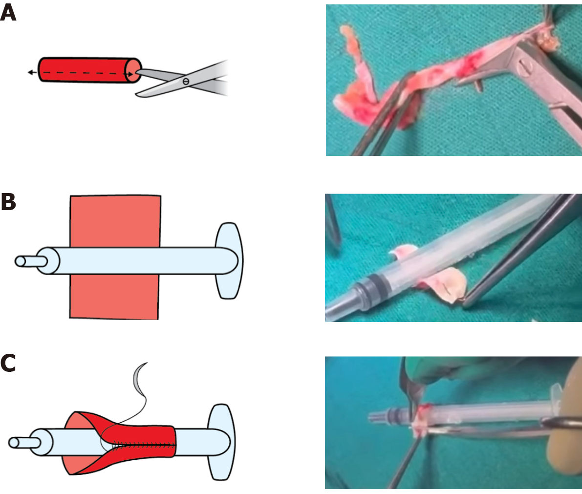
Figure 4 The recipient’s inferior epigastric artery was carefully dissected, opened longitudinally, and molded over a syringe to preserve its cylindric shape, thereby creating a vascular cuff for extension and anastomosis with the donor’s renal artery.
A: Longitudinal incision of the epigastric artery; B: Molding of the epigastric artery transversally over the syringe; C: Creation of the vascular cuff using a continuous suture.
- Citation: Lekehal B, Ait Youssef N, Lekehal M, Bakkali T, Jdar A, Bounssir A. Inferior epigastric artery cuff interposition for short renal artery in living-donor kidney transplantation: A case report and review of literature. World J Transplant 2025; 15(4): 109968
- URL: https://www.wjgnet.com/2220-3230/full/v15/i4/109968.htm
- DOI: https://dx.doi.org/10.5500/wjt.v15.i4.109968