
















©The Author(s) 2024.
World J Transplant. Jun 18, 2024; 14(2): 92137
Published online Jun 18, 2024. doi: 10.5500/wjt.v14.i2.92137
Published online Jun 18, 2024. doi: 10.5500/wjt.v14.i2.92137
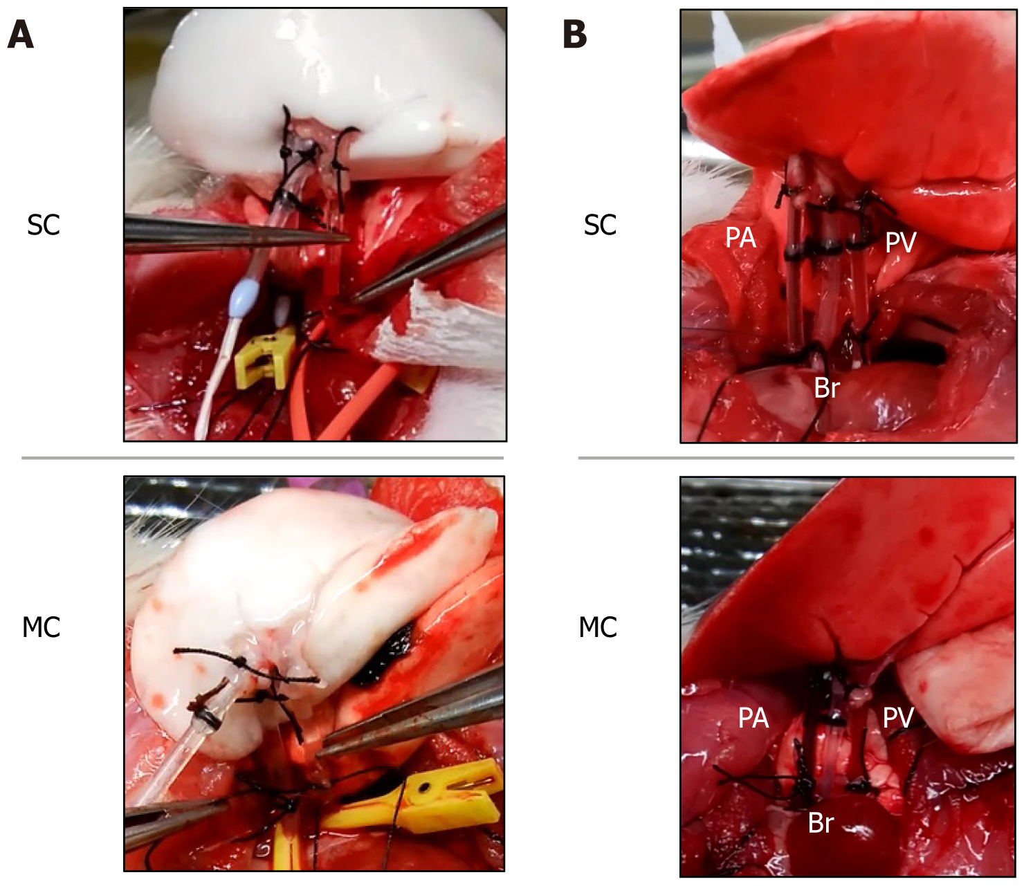
Figure 3 Recipient operation.
A: Pulmonary vein anastomosis using a cannula; B: After lung graft revascularization, venous blood perfusion is visible in the pulmonary artery, and arterial blood perfusion is visible in the pulmonary artery. In the modified cannula group, the grafted lung is located closer to the pulmonary hilum compared with the shunt cannul group. PA: Pulmonary artery; PV: Pulmonary vein; Br: Bronchus; MC: Modified cannula; SC: Shunt cannula.
- Citation: Takata M, Tanaka Y, Saito D, Yoshida S, Matsumoto I. Hyperacute experimental model of rat lung transplantation using a coronary shunt cannula. World J Transplant 2024; 14(2): 92137
- URL: https://www.wjgnet.com/2220-3230/full/v14/i2/92137.htm
- DOI: https://dx.doi.org/10.5500/wjt.v14.i2.92137