Copyright
©The Author(s) 2021.
World J Transplant. Jun 18, 2021; 11(6): 244-253
Published online Jun 18, 2021. doi: 10.5500/wjt.v11.i6.244
Published online Jun 18, 2021. doi: 10.5500/wjt.v11.i6.244
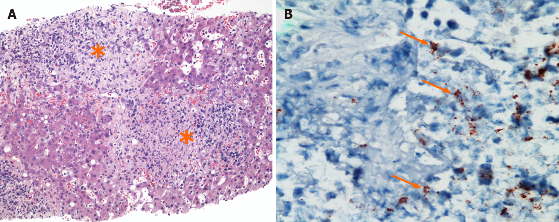
Figure 1 Histopathology of liver biopsy from case 1.
A: Photomicrograph of liver parenchyma with non-necrotizing granulomas (asterisks) with histiocytes, lymphoplasmacytic inflammatory cells, and neutrophils. (hematoxylin and eosin, 200 ×); B: Bartonella henselae immunohistochemical stain highlights individual and clustered coccobacilli (arrows) (400 ×).
- Citation: Pischel L, Radcliffe C, Vilchez GA, Charifa A, Zhang XC, Grant M. Bartonellosis in transplant recipients: A retrospective single center experience. World J Transplant 2021; 11(6): 244-253
- URL: https://www.wjgnet.com/2220-3230/full/v11/i6/244.htm
- DOI: https://dx.doi.org/10.5500/wjt.v11.i6.244


