
















©The Author(s) 2021.
World J Transplant. Jun 18, 2021; 11(6): 231-243
Published online Jun 18, 2021. doi: 10.5500/wjt.v11.i6.231
Published online Jun 18, 2021. doi: 10.5500/wjt.v11.i6.231
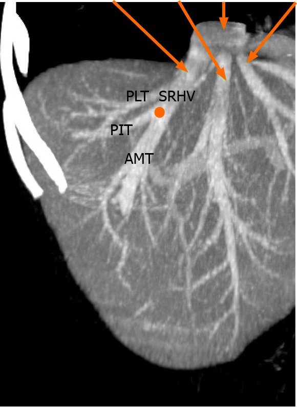
Figure 4 Variations of the right hepatic vein.
Coronal view of reconstructed computed tomography images demonstrating a proximal venous confluence (orange) that receives the posteroinferior tributaries (PITs) and anteromedial tributaries (AMTs) before continuing cephalad as the superior right hepatic vein (SRHV). The consistent posterolateral tributary (PLT) from segment VII is also seen.
- Citation: Cawich SO, Naraynsingh V, Pearce NW, Deshpande RR, Rampersad R, Gardner MT, Mohammed F, Dindial R, Barrow TA. Surgical relevance of anatomic variations of the right hepatic vein. World J Transplant 2021; 11(6): 231-243
- URL: https://www.wjgnet.com/2220-3230/full/v11/i6/231.htm
- DOI: https://dx.doi.org/10.5500/wjt.v11.i6.231