Copyright
©The Author(s) 2016.
World J Transplant. Mar 24, 2016; 6(1): 215-219
Published online Mar 24, 2016. doi: 10.5500/wjt.v6.i1.215
Published online Mar 24, 2016. doi: 10.5500/wjt.v6.i1.215
Figure 1 Computed tomography of chest.
A: Day 40. Note a well circumscribed, round pulmonary nodule involving the right lower lobe. Transbronchial biopsy was obtained from this site 40 d earlier; B: Day 90. Note the total resolution of the right lower lobe nodule.
Figure 2 Postroanterior and lateral views of the chest.
A: Day 40. Note a well circumscribed, round pulmonary nodule involving the RLL, 2 cm in diameter. Transbronchial biopsy was obtained from this site 40 d earlier; B: Day 90. Note the total resolution of the RLL nodule. RLL: Right lower lobe.
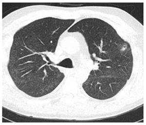
Figure 3 Computed tomography of chest revealing a cavitating lung nodule involving lingual.
A transbronchial biopsy was obtained from the site 21 d earlier.
- Citation: Mehta AC, Wang J, Abuqayyas S, Garcha P, Lane CR, Tsuang W, Budev M, Akindipe O. New Nodule-Newer Etiology. World J Transplant 2016; 6(1): 215-219
- URL: https://www.wjgnet.com/2220-3230/full/v6/i1/215.htm
- DOI: https://dx.doi.org/10.5500/wjt.v6.i1.215


