Copyright
©The Author(s) 2015.
World J Psychiatr. Jun 22, 2015; 5(2): 193-209
Published online Jun 22, 2015. doi: 10.5498/wjp.v5.i2.193
Published online Jun 22, 2015. doi: 10.5498/wjp.v5.i2.193
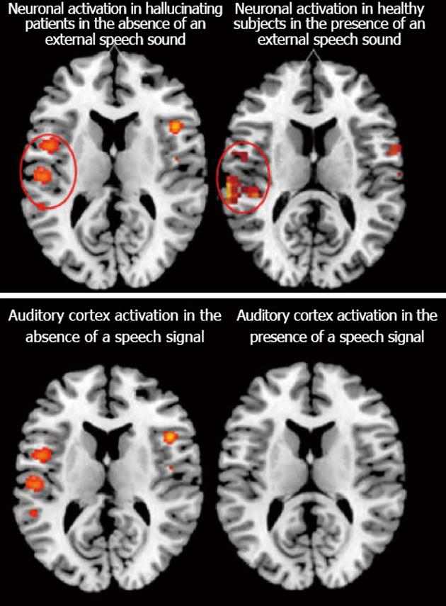
Figure 3 Functional imaging results for hallucinating patients based on the meta-analysis done by Kompus et al[24] which show activation in the absence of an external auditory stimulus (upper left panel) compared with healthy controls in the presence of an external auditory stimulus (upper right panel, data from van den Noort et al[117]).
The lower left panel shows the same activation as in the upper left panel, spontaneous activation in hallucinating patients in the absence of an external auditory stimulus, but now compared with absence of activation in hallucinating patients in the presence of an auditory stimulus. Color-coded areas indicate significantly activated brain regions during active hallucinations and task-processing. Reprinted and redrawn with permission from the authors and the publishers.
- Citation: Hugdahl K. Auditory hallucinations: A review of the ERC “VOICE” project. World J Psychiatr 2015; 5(2): 193-209
- URL: https://www.wjgnet.com/2220-3206/full/v5/i2/193.htm
- DOI: https://dx.doi.org/10.5498/wjp.v5.i2.193


