Copyright
©The Author(s) 2015.
World J Psychiatr. Mar 22, 2015; 5(1): 15-34
Published online Mar 22, 2015. doi: 10.5498/wjp.v5.i1.15
Published online Mar 22, 2015. doi: 10.5498/wjp.v5.i1.15
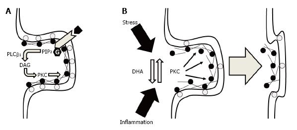
Figure 1 Potential ultrastructural mechanisms by which membrane DHA deficits could lead to a loss in synaptic connectivity.
A: Under normal physiological conditions, synaptic membrane phosphatidylserine (gray circles) bind MARCKS (black circles) at the membrane which also cross-links and tethers F-actin to support dendritic spine cytoskeletal structural stability. Binding of MARCKS with membrane phosphatidylserine also inhibits phospholipase Cβ1-mediated hydrolysis of phosphatidylinositol 4,5-bisphosphate (PIP2) into diacylglycerol (DAG) which increases PKC activity. PKC-mediated phosphorylation of MARCKS reduces the tensile strength of the F-actin cytoskeleton leading to the eventual collapse of the spine; B: Under conditions of membrane DHA deficits, reductions in membrane phosphatidylserine and membrane-bound MARCKS increase PKC activity and destabilizes the F-actin cytoskeleton leading to spine collapse. Elevated PKC activity secondary to membrane DHA deficits may also reduce the resilience of dendritic spines to other pathophysiological factors including chronic stress or inflammation. DHA: Docosahexaenoic acid; PKC: Protein kinase C; MARCKS: Myristoylated alanine-rich C kinase substrate.
- Citation: McNamara RK, Vannest JJ, Valentine CJ. Role of perinatal long-chain omega-3 fatty acids in cortical circuit maturation: Mechanisms and implications for psychopathology. World J Psychiatr 2015; 5(1): 15-34
- URL: https://www.wjgnet.com/2220-3206/full/v5/i1/15.htm
- DOI: https://dx.doi.org/10.5498/wjp.v5.i1.15


