
















Copyright: ©Author(s) 2026.
World J Psychiatry. Jul 19, 2026; 16(7): 117775
Published online Jul 19, 2026. doi: 10.5498/wjp.117775
Published online Jul 19, 2026. doi: 10.5498/wjp.117775
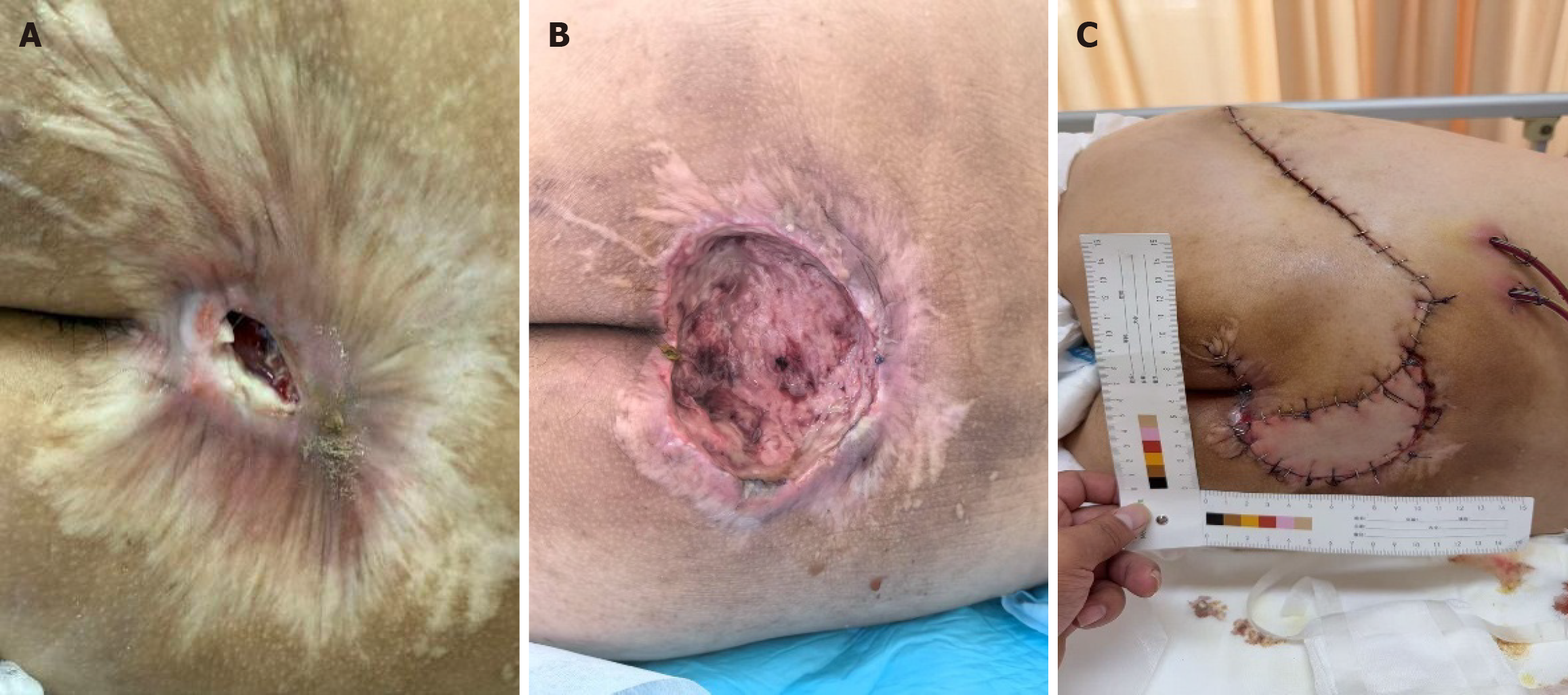
Figure 1 The pressure ulcer repair process in the control group patient.
A: The sacrococcygeal area presented with a 7 cm × 9 cm area of scar contracture. Within this, a central 6 cm × 7 cm region exhibited erythema and swelling, featuring a 3 cm × 3 cm open wound that extended to the depth of the sacrum and coccyx. The surface of the sacrococcygeal bone appeared black and was covered with purulent slough; B: Postoperative view following debridement of the sacrococcygeal osteomyelitic lesion, muscle debridement for ulcer repair, and application of a vacuum sealing drainage system; C: Postoperative view after a gluteus maximus myocutaneous flap transfer and skin grafting.
- Citation: Xing J, Jia YJ, Xin JQ, Liu JY, Cui Y, Zhang HW. Watson caring theory-based encouragement interventions for pain and anxiety in stage III-IV pressure ulcer patients. World J Psychiatry 2026; 16(7): 117775
- URL: https://www.wjgnet.com/2220-3206/full/v16/i7/117775.htm
- DOI: https://dx.doi.org/10.5498/wjp.117775