
















Copyright: ©Author(s) 2026.
World J Psychiatry. Jun 19, 2026; 16(6): 117066
Published online Jun 19, 2026. doi: 10.5498/wjp.v16.i6.117066
Published online Jun 19, 2026. doi: 10.5498/wjp.v16.i6.117066
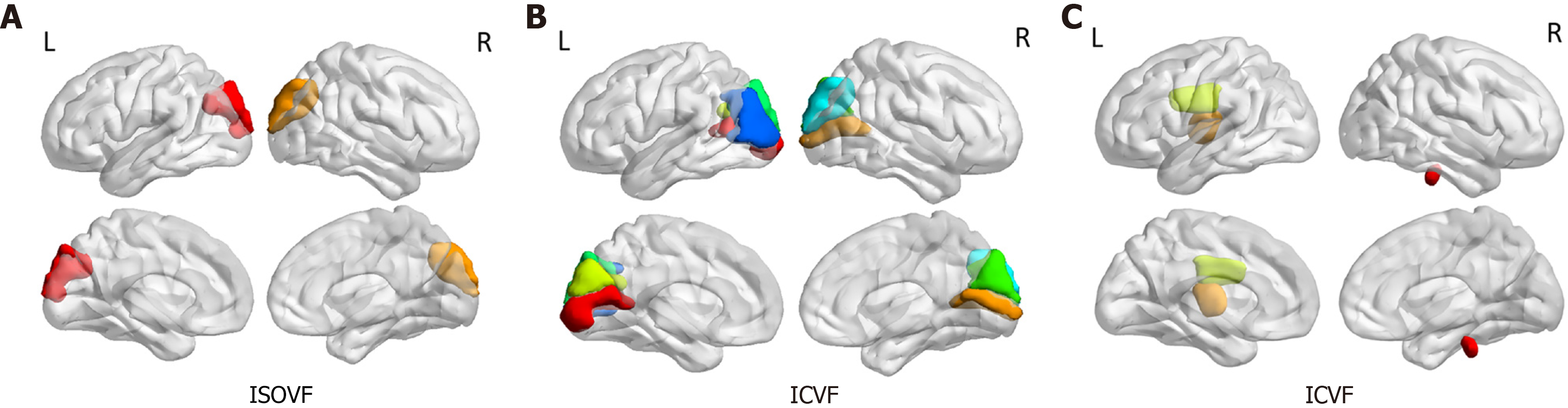
Figure 2 Brain regions showing significant differences in volume fraction of the isotropically diffusing water and intracellular volume fraction of gray/white matter values between the circadian rhythm disruption and control groups.
A: Significantly higher volume fraction of the isotropically diffusing water values in gray matter were found in the circadian rhythm disruption (CRD) group compared with the control group in the left and right superior occipital gyrus; B: Values of intracellular volume fraction (ICVF) in gray matter were significantly higher in the CRD group compared to the control group in the following brain regions: Left calcarine fissure and surrounding cortex, right calcarine fissure and surrounding cortex, left cuneus, right cuneus, left superior occipital gyrus, right superior occipital gyrus, and left middle occipital gyrus; C: ICVF levels in white matter were also significantly higher in the CRD group compared to the control group, specifically in the pontine crossing tract, left posterior limb of the internal capsule, and left superior coronal radiata. L: Left; R: Right; ICVF: Intracellular volume fraction; ISOVF: Volume fraction of the isotropically diffusing water.
- Citation: Zhang F, Huang WY, Zhang L, Huang ZR, Zhao W, Yu YQ. Circadian rhythm disruption alters gray and white matter microstructure: Higher-order diffusion analysis coupled with hormonal/inflammatory biomarkers. World J Psychiatry 2026; 16(6): 117066
- URL: https://www.wjgnet.com/2220-3206/full/v16/i6/117066.htm
- DOI: https://dx.doi.org/10.5498/wjp.v16.i6.117066