
















Copyright: ©Author(s) 2026.
World J Psychiatry. Jun 19, 2026; 16(6): 115996
Published online Jun 19, 2026. doi: 10.5498/wjp.v16.i6.115996
Published online Jun 19, 2026. doi: 10.5498/wjp.v16.i6.115996
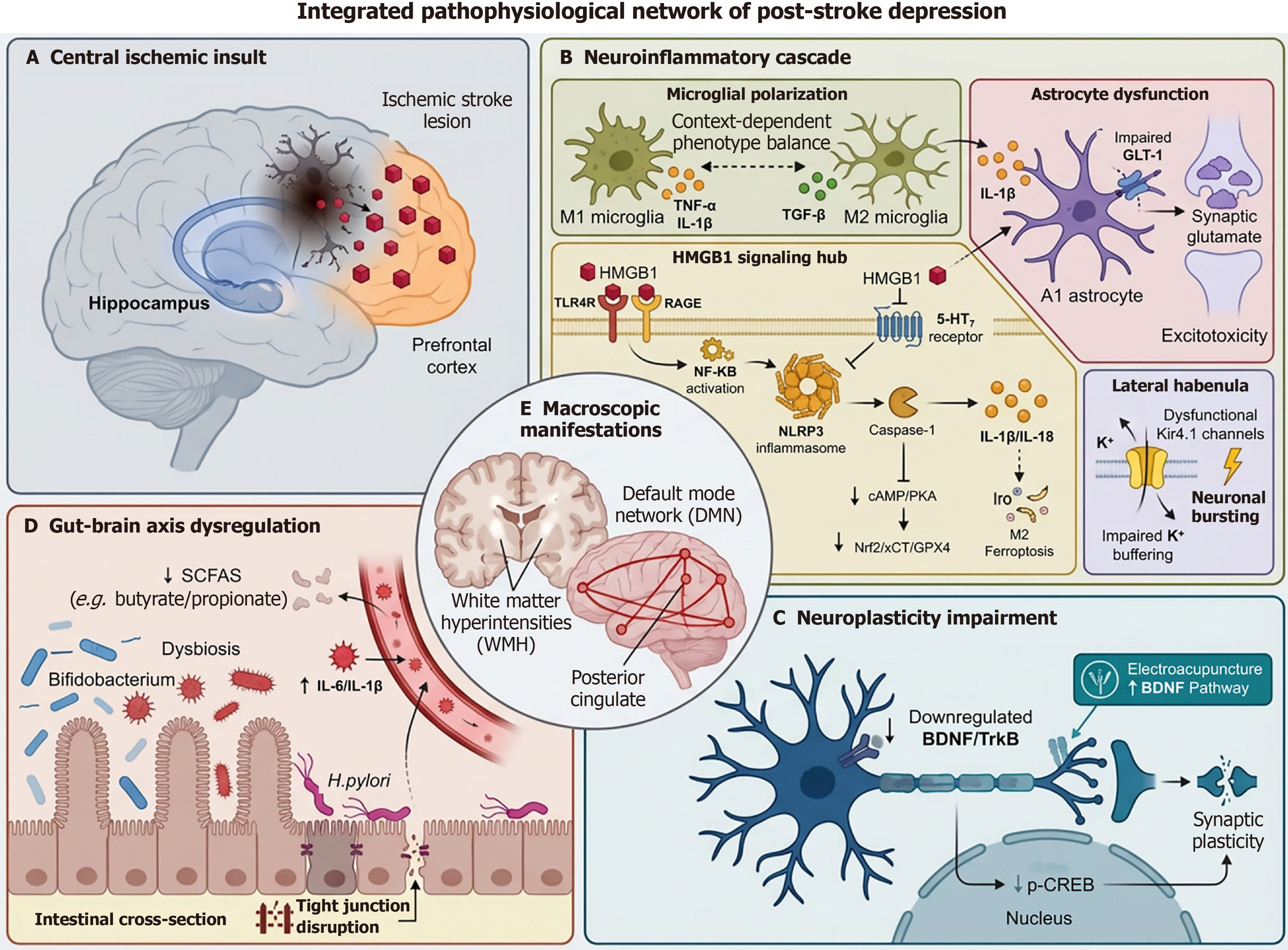
Figure 4 Integrated pathophysiological network of post-stroke depression.
A: Central ischemic insult. Focal brain ischemia, depicted in the hippocampus and prefrontal cortex, causes neuronal necrosis and serves as the primary trigger, releasing damage signals that initiate downstream pathological cascades; B: Neuroinflammatory Cascade. high-mobility group box 1 emerges as a pivotal hub, released from damaged neurons to activate microglia via RAGE/TLR4, driving NF-κB and NLRP3 inflammasome signaling. This sustains a cycle of M1 microglial activation and A1 astrocytic reactivity, with the latter contributing to excitotoxicity via impaired GLT-1 function; C: Neuroplasticity Impairment. A hallmark of post-stroke depression (PSD) is the downregulation of the BDNF/TrkB/CREB signaling pathway, crucial for neuronal survival and synaptic integrity. This impairment directly links cellular stress to the failure of neural circuit adaptation; D: Gut-brain axis dysregulation. Post-stroke gut dysbiosis-characterized by a loss of beneficial short-chain fatty acid-producing bacteria and an increase in opportunistic pathogens-compromises intestinal barrier function, amplifying systemic inflammation that can exacerbate central neuroinflammation; E: Macroscopic network manifestations. The molecular and cellular perturbations culminate in structural changes (e.g., white matter hyperintensities) and functional default mode network dysfunction, particularly hyperconnectivity in hubs like the posterior cingulate cortex, which underlies cognitive-emotional deficits in PSD.
- Citation: Gu JX, Liu CQ, Chen GX, Yao T, Sun ZX, Wang Y. Post-stroke depression update 2025: Mechanisms, prediction, and management. World J Psychiatry 2026; 16(6): 115996
- URL: https://www.wjgnet.com/2220-3206/full/v16/i6/115996.htm
- DOI: https://dx.doi.org/10.5498/wjp.v16.i6.115996