
















Copyright: ©Author(s) 2026.
World J Psychiatry. May 19, 2026; 16(5): 114524
Published online May 19, 2026. doi: 10.5498/wjp.v16.i5.114524
Published online May 19, 2026. doi: 10.5498/wjp.v16.i5.114524
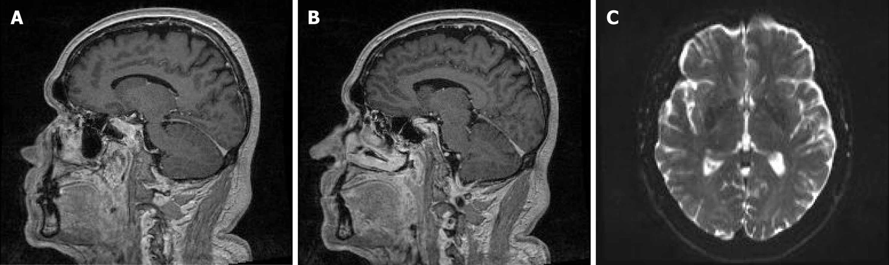
Figure 1 Magnetic resonance imaging brain and neck normal with a well-defined, homogeneously hyperintense mass on both T1- and T2-weighted sequences, which is fully suppressed on fatsaturated images-consistent with mature adipose tissue confined to the sub
- Citation: Stip E. Adjustment disorder triggered by a unique and pervasive urban stressor, traffic cones: A case report. World J Psychiatry 2026; 16(5): 114524
- URL: https://www.wjgnet.com/2220-3206/full/v16/i5/114524.htm
- DOI: https://dx.doi.org/10.5498/wjp.v16.i5.114524