
















©The Author(s) 2026.
World J Psychiatry. Jan 19, 2026; 16(1): 113230
Published online Jan 19, 2026. doi: 10.5498/wjp.v16.i1.113230
Published online Jan 19, 2026. doi: 10.5498/wjp.v16.i1.113230
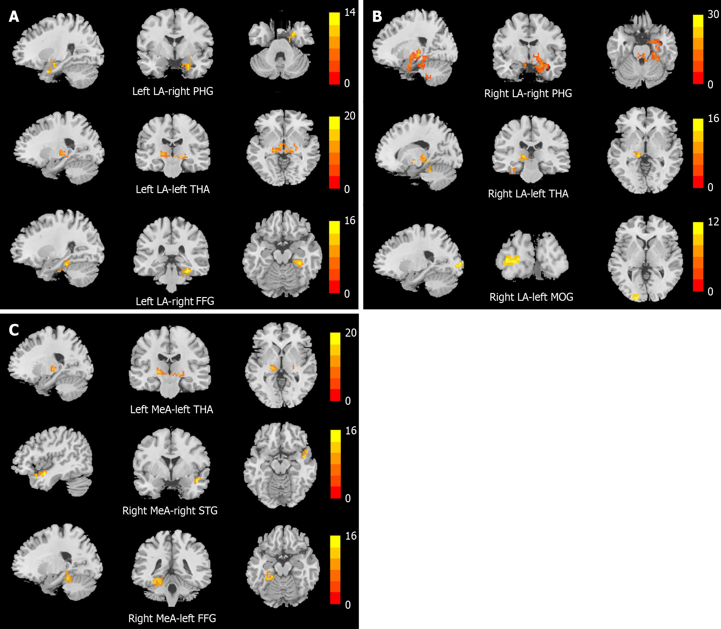
Figure 2 The brain regions with significant differences in resting-state functional connectivity of the amygdala subregions among the three groups.
A: Brain regions with significant differences in resting-state functional connectivity (rsFC) of the left lateral amygdala (LA); B: Brain regions with significant differences in rsFC of the right LA; C: Brain regions with significant differences in rsFC of the medial amygdala. P < 0.001 for voxel level and P < 0.05 for cluster level, gaussian random field correction. The colored bars represent the F values. FFG: Fusiform gyrus; LA: Lateral amygdala; MeA: Medial amygdala; MOG: Middle occipital gyrus; PHG: Parahippocampal gyrus; STG: Superior temporal gyrus; THA: Thalamus.
- Citation: Cheng SC, Wang YM, Li YT, Yao QN, Huang XL, Ji J, Zhang XB, Sun HY. Aberrant resting-state functional connectivity in amygdala subregions among adolescents with depression and suicide attempts. World J Psychiatry 2026; 16(1): 113230
- URL: https://www.wjgnet.com/2220-3206/full/v16/i1/113230.htm
- DOI: https://dx.doi.org/10.5498/wjp.v16.i1.113230