
















©The Author(s) 2025.
World J Psychiatry. Dec 19, 2025; 15(12): 111754
Published online Dec 19, 2025. doi: 10.5498/wjp.v15.i12.111754
Published online Dec 19, 2025. doi: 10.5498/wjp.v15.i12.111754
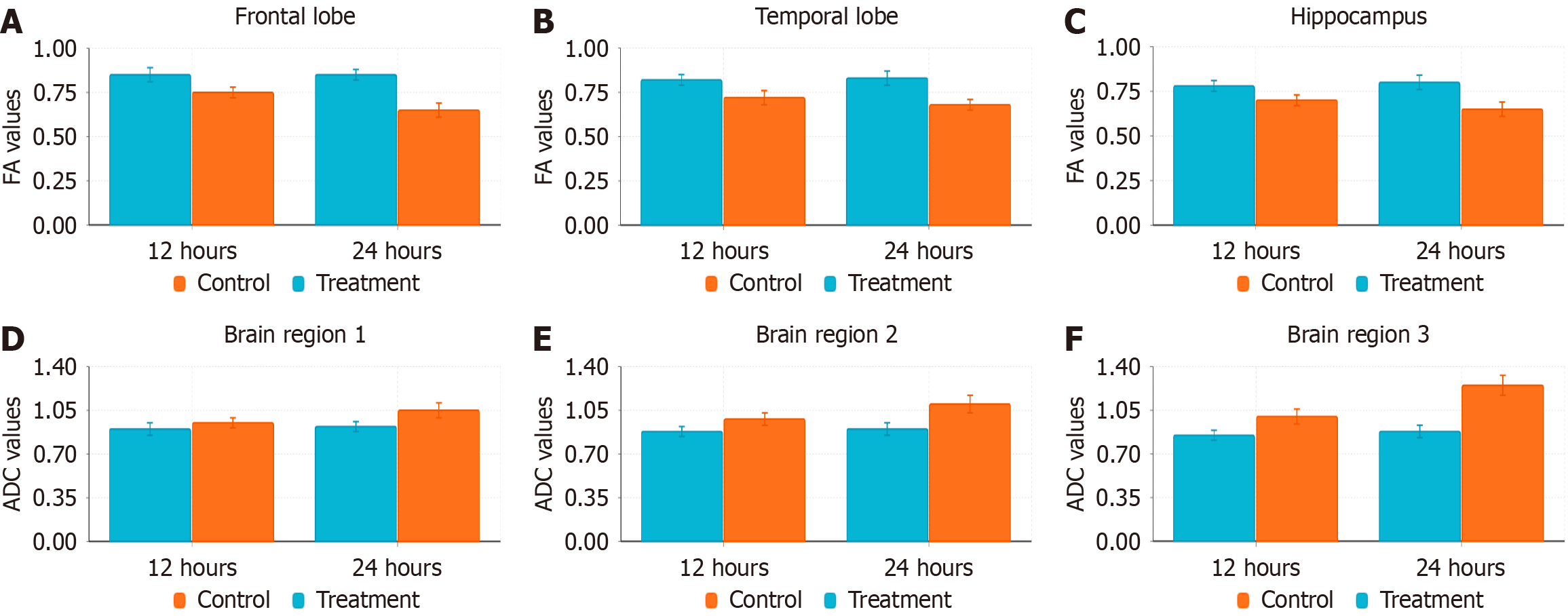
Figure 2 Changes in diffusion tensor imaging parameters in the frontal lobe, temporal lobe, and hippocampus of rats after subarachnoid hemorrhage.
A-C: Fractional anisotropy values in the frontal lobe (A), temporal lobe (B), and hippocampus (C) gradually decreased after subarachnoid hemorrhage (SAH), reflecting damage to white matter fiber integrity; D-F: Apparent diffusion coefficient values in the frontal lobe (D), temporal lobe (E), and hippocampus (F) gradually increased after SAH, suggesting cellular edema and tissue damage. Compared with the control group (cyan bars), the SAH group (orange bars) showed the most significant changes in diffusion tensor imaging parameters in the hippocampus, especially at the 24 hours time point. Data are presented as mean ± SD. FA: Fractional anisotropy; ADC: Apparent diffusion coefficient.
- Citation: Qin L, Wang K, Jiang LP, Xiao Z, Luo S. Correlation between anxiety-depression disorders and brain structural connectivity abnormalities after subarachnoid hemorrhage. World J Psychiatry 2025; 15(12): 111754
- URL: https://www.wjgnet.com/2220-3206/full/v15/i12/111754.htm
- DOI: https://dx.doi.org/10.5498/wjp.v15.i12.111754