
















©The Author(s) 2025.
World J Psychiatry. Dec 19, 2025; 15(12): 110916
Published online Dec 19, 2025. doi: 10.5498/wjp.v15.i12.110916
Published online Dec 19, 2025. doi: 10.5498/wjp.v15.i12.110916
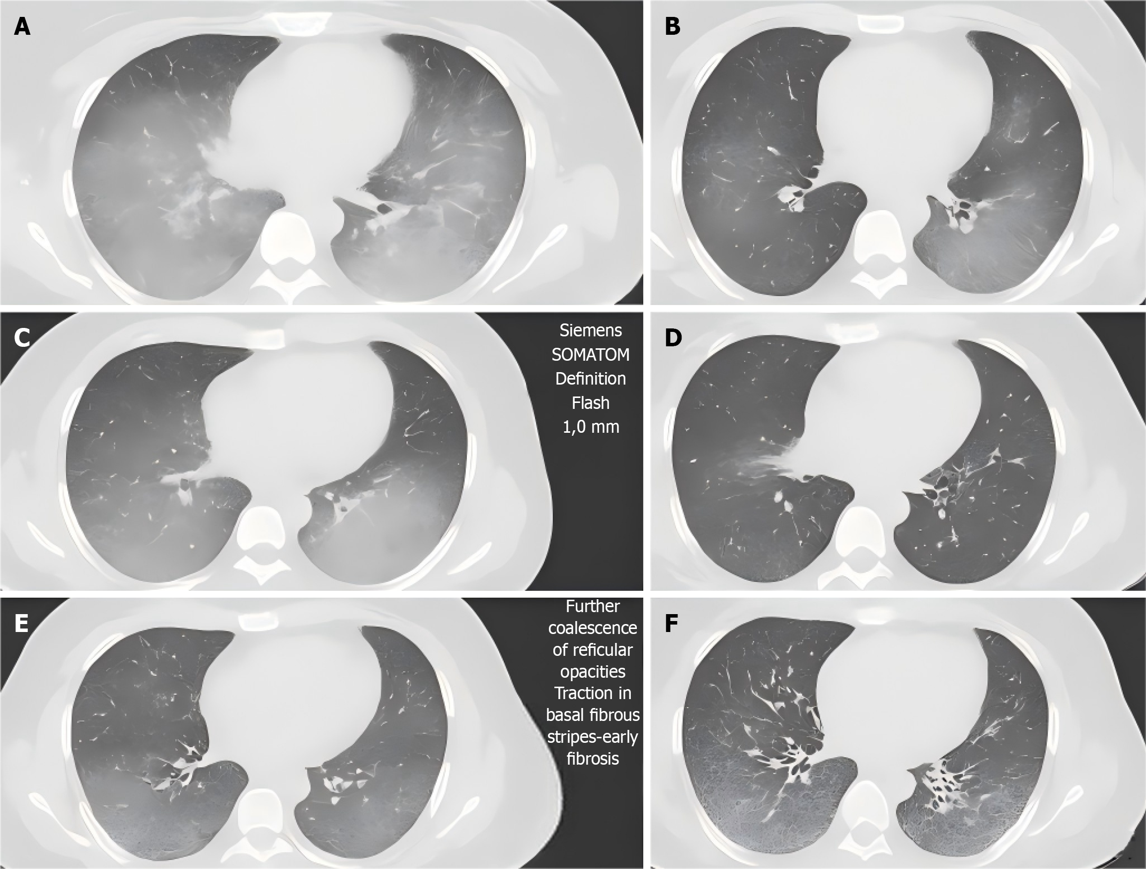
Figure 1 Detection of high-resolution computed tomography imaging indicators in typical cases.
A and B: Diffuse grid shadow with ground-glass shadow in middle and lower fields of both lungs, and subpleural distribution was dominant (at enrollment); C and D: The ground-glass opacity was partially absorbed, but fibrous bands were increased in the basal segment, suggesting early fibrosis (6th month); E and F: Shows further confluency of the grid shadows, with the formation of traction branches and the appearance of honeycomb-like changes with continuous progression of fibrosis (12th month). Scan parameters: Siemens Somatom Definition Flash, 1.0 mm layer thickness, 120 kVp, 200-250 mAs. Double blind evaluation of quantitative lesion extent.
- Citation: Zhu ZJ, Liu KL, Qu HR. Relationship between anxiety and depression, dyspnea severity, and pulmonary function in connective tissue disease-associated interstitial lung disease. World J Psychiatry 2025; 15(12): 110916
- URL: https://www.wjgnet.com/2220-3206/full/v15/i12/110916.htm
- DOI: https://dx.doi.org/10.5498/wjp.v15.i12.110916