
















©The Author(s) 2025.
World J Psychiatry. Dec 19, 2025; 15(12): 108867
Published online Dec 19, 2025. doi: 10.5498/wjp.v15.i12.108867
Published online Dec 19, 2025. doi: 10.5498/wjp.v15.i12.108867
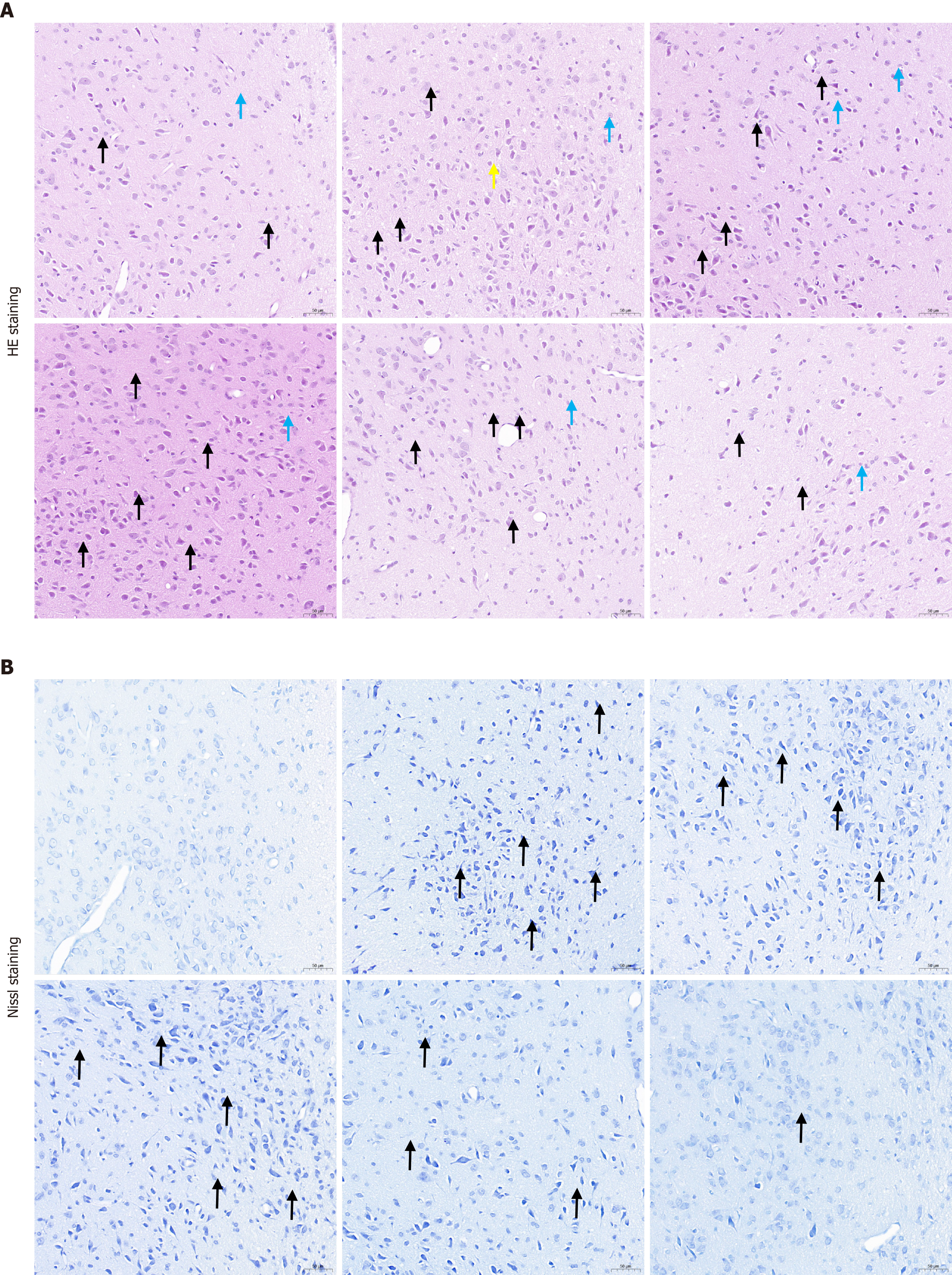
Figure 4 Histological evaluation of the hypothalamus using haematoxylin and eosin staining and Nissl staining.
A: Haematoxylin and eosin staining: Hypothalamic neurons exhibit shrunken and deeply stained cell bodies (indicated by black arrows), increased glial cell density (blue arrows), and occasional neurophagocytosis (yellow arrows); B: Nissl staining: Neuronal cell bodies are shrunken and deeply stained (black arrows), with loss of Nissl bodies in the cytoplasm. HE: Haematoxylin and eosin.
- Citation: Li RT, Lan BJ, Xiao ZY, Shi QH, Chen XY, Li F. Sini-Suanzaoren decoction regulates mitochondrial biogenesis mediated by MT-SIRT1 in the treatment of insomnia rats. World J Psychiatry 2025; 15(12): 108867
- URL: https://www.wjgnet.com/2220-3206/full/v15/i12/108867.htm
- DOI: https://dx.doi.org/10.5498/wjp.v15.i12.108867