Copyright
©The Author(s) 2015.
World J Clin Infect Dis. Nov 25, 2015; 5(4): 67-76
Published online Nov 25, 2015. doi: 10.5495/wjcid.v5.i4.67
Published online Nov 25, 2015. doi: 10.5495/wjcid.v5.i4.67
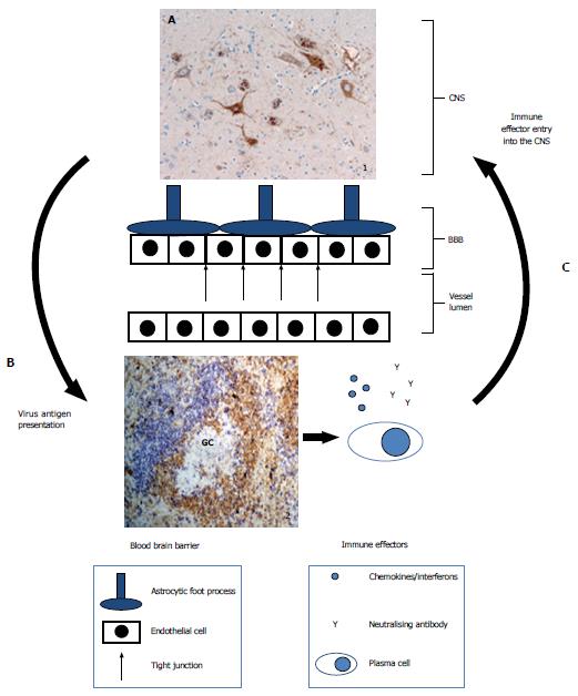
Figure 2 Model showing the movement of rabies virus antigen from the central nervous system to lymphoid tissue and then immune effectors (chemokines, antibodies and plasma cells) return to the central nervous system.
Inset 1 shows a section of RABV infected brain stained with anti-nucleoprotein antibodies. Brown staining shows accumulation of nucleoprotein within neuronal cells. Inset 2 shows a murine spleen section stained with anti-CD3 for T cells (blue staining) and anti-IgD for naïve B cells (brown staining). The location of a GC is indicated. The arrows indicate the movement of antigen to lymphoid tissue and immune effectors returning to the CNS. The exact route of these is uncertain in the context of RABV infection. Letters signify areas for future research: A: Identification of therapeutics that restrict the replication and spread of RABV between neurons. Critically, such antiviral strategies must work within the CNS and without toxicity; B: Investigate the mechanisms by which RABV antigens reach antigen presenting cells either in situ or through exit of the CNS. Therapies that accelerate this process should assist the development of immune responses to infection; C: Development of methods that improves access of antivirals and immunes effectors (cytokines, antibodies and cells) into the CNS. RABV: Rabies virus; CNS: Central nervous system; GC: Germinal centre.
- Citation: Johnson N, Cunningham AF. Interplay between rabies virus and the mammalian immune system. World J Clin Infect Dis 2015; 5(4): 67-76
- URL: https://www.wjgnet.com/2220-3176/full/v5/i4/67.htm
- DOI: https://dx.doi.org/10.5495/wjcid.v5.i4.67


