
















Copyright: ©Author(s) 2026.
World J Exp Med. Sep 20, 2026; 16(3): 126036
Published online Sep 20, 2026. doi: 10.5493/wjem.126036
Published online Sep 20, 2026. doi: 10.5493/wjem.126036
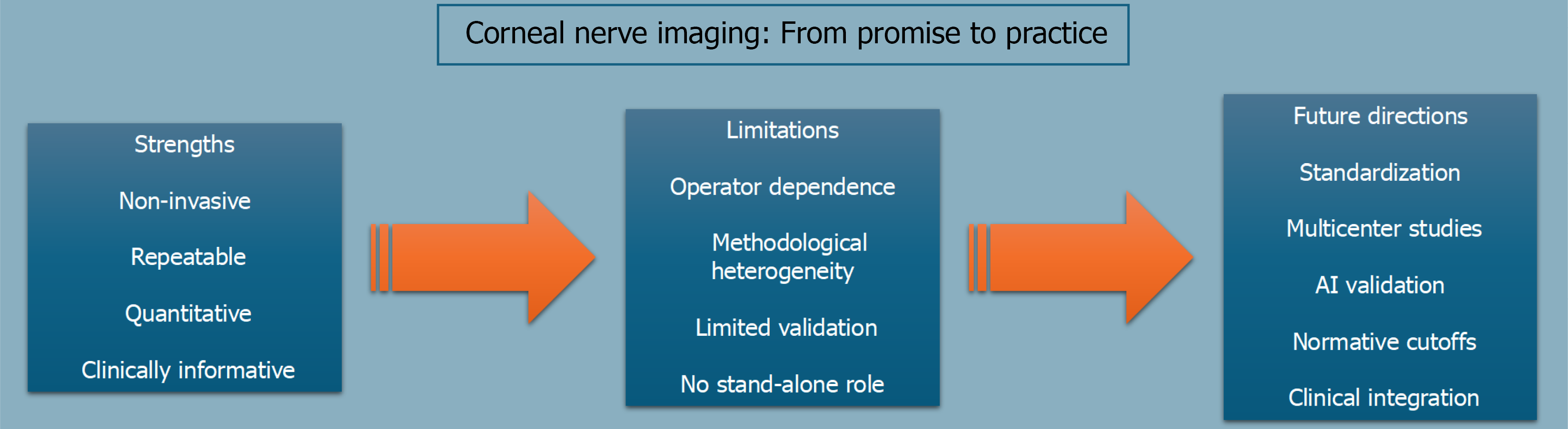
Figure 2 Corneal nerve imaging: From promise to practice.
Corneal confocal microscopy offers several practical strengths for evaluating small-fiber damage, including noninvasiveness, repeatability, quantification, and clinical utility. However, wider clinical implementation is still limited by operator dependence, methodological heterogeneity, incomplete validation, and the lack of a stand-alone diagnostic role. Future progress will depend on stronger standardization, multicenter studies, artificial intelligence validation, normative cutoffs, and closer integration into clinical practice.
- Citation: Capobianco M, Cappellani F, Enaholo E, Zeppieri M. Corneal nerve imaging in systemic neuropathy: A minireview of experimental and precision medicine applications. World J Exp Med 2026; 16(3): 126036
- URL: https://www.wjgnet.com/2220-315x/full/v16/i3/126036.htm
- DOI: https://dx.doi.org/10.5493/wjem.126036