
















Copyright: ©Author(s) 2026.
World J Exp Med. Sep 20, 2026; 16(3): 126036
Published online Sep 20, 2026. doi: 10.5493/wjem.126036
Published online Sep 20, 2026. doi: 10.5493/wjem.126036
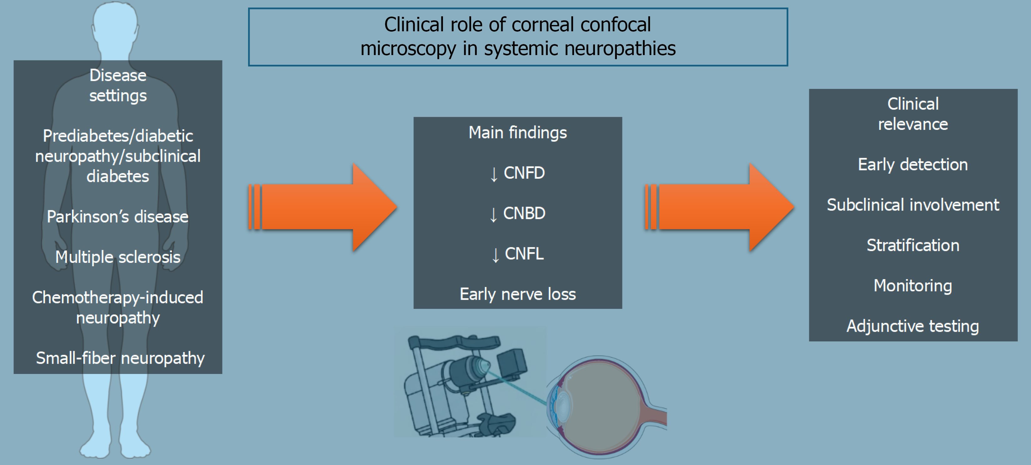
Figure 1 Clinical role of corneal confocal microscopy in systemic neuropathies.
Corneal confocal microscopy has been investigated in several systemic and neurologic conditions, including prediabetes and diabetic neuropathy, Parkinson’s disease, multiple sclerosis, chemotherapy-induced neuropathy, and small-fiber neuropathy. Across these settings, the most consistent findings are reductions in corneal nerve fiber density, branch density, and length, consistent with early corneal nerve loss. These alterations may support early detection of small-fiber damage, identification of subclinical involvement, patient stratification, longitudinal monitoring, and adjunctive assessment of neuropathy. These findings primarily describe associations observed at the group level; no disease-specific corneal confocal microscopy pattern or universally validated threshold currently establishes systemic neuropathy in an individual patient. Image(s) provided by Servier Medical Art (https://smart.servier.com), licensed under CC BY 4.0 (https://creativecommons.org/Licenses/by/4.0/). CNFD: Corneal nerve fiber density; CNBD: Corneal nerve branch density; CNFL: Corneal nerve fiber length.
- Citation: Capobianco M, Cappellani F, Enaholo E, Zeppieri M. Corneal nerve imaging in systemic neuropathy: A minireview of experimental and precision medicine applications. World J Exp Med 2026; 16(3): 126036
- URL: https://www.wjgnet.com/2220-315x/full/v16/i3/126036.htm
- DOI: https://dx.doi.org/10.5493/wjem.126036