
















Copyright: ©Author(s) 2026.
World J Exp Med. Jun 20, 2026; 16(2): 118828
Published online Jun 20, 2026. doi: 10.5493/wjem.118828
Published online Jun 20, 2026. doi: 10.5493/wjem.118828
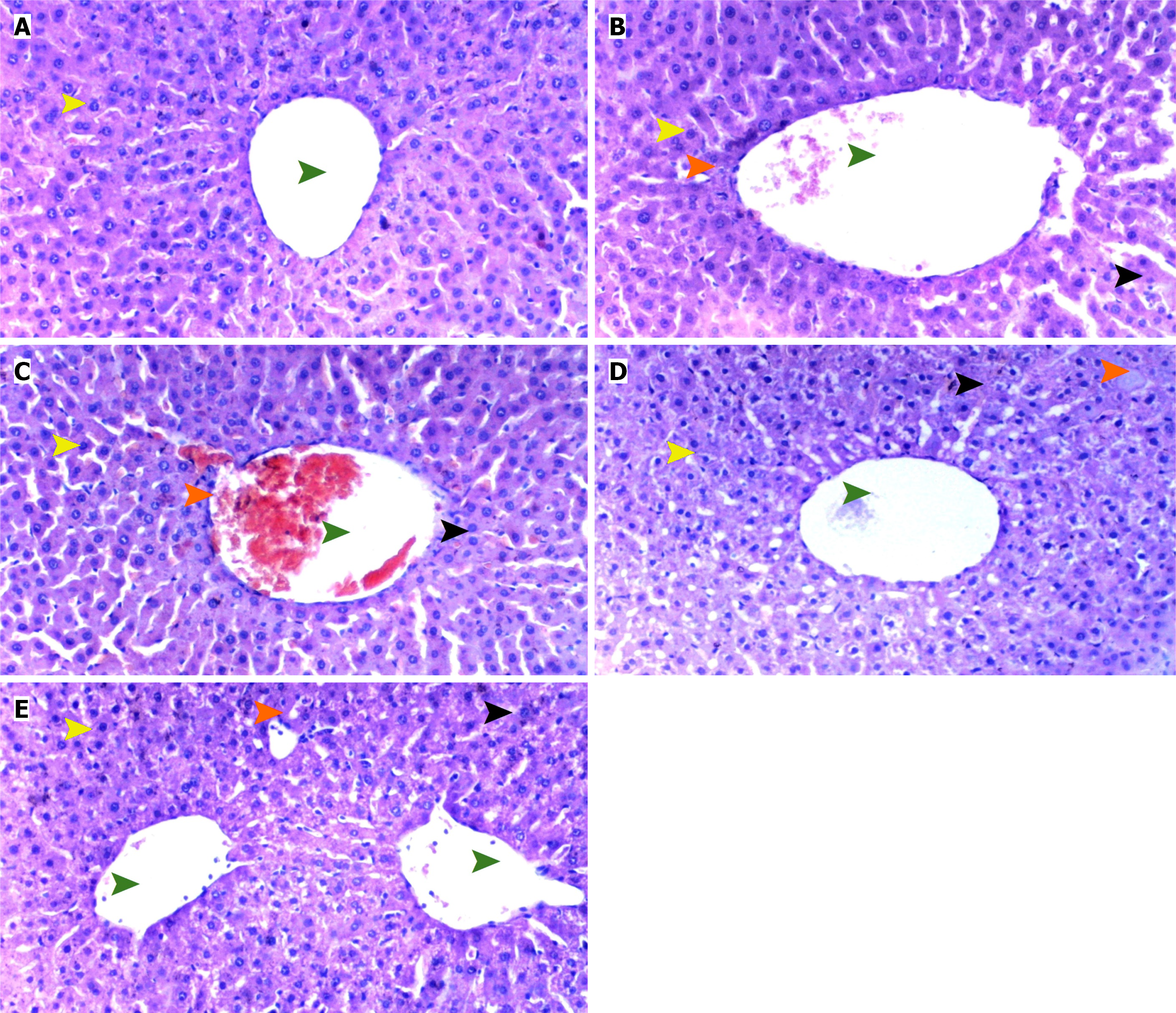
Figure 1 Histopathological findings in liver tissue.
A: Normal liver tissue, control group, hematoxylin and eosin staining (HE) × 100; B: Ischemia-reperfusion group, HE × 100; C: Ischemia-reperfusion rectal ozone group, HE × 100; D: Ischemia-reperfusion intrathecal ozone group, HE × 100; E: Ischemia-reperfusion intraperitoneal ozone group, HE × 100, scale bar 50 μm. Green arrow: Vena centralis; orange arrow: Degeneration; black arrow: Inflammation; yellow arrow: Hepatocytes.
- Citation: Şengel N, Köksal Z, Küçük A, Sezen ŞC, Güneş I, Atlı M, Kip G, Gökgöz Acar S, Özer A, Kavutçu M, Arslan M. Comparison of ozone administration routes on liver, lung, and brain damage after spinal cord ischemia-reperfusion injury. World J Exp Med 2026; 16(2): 118828
- URL: https://www.wjgnet.com/2220-315x/full/v16/i2/118828.htm
- DOI: https://dx.doi.org/10.5493/wjem.118828