
















Copyright: ©Author(s) 2026.
World J Exp Med. Mar 20, 2026; 16(1): 115508
Published online Mar 20, 2026. doi: 10.5493/wjem.v16.i1.115508
Published online Mar 20, 2026. doi: 10.5493/wjem.v16.i1.115508
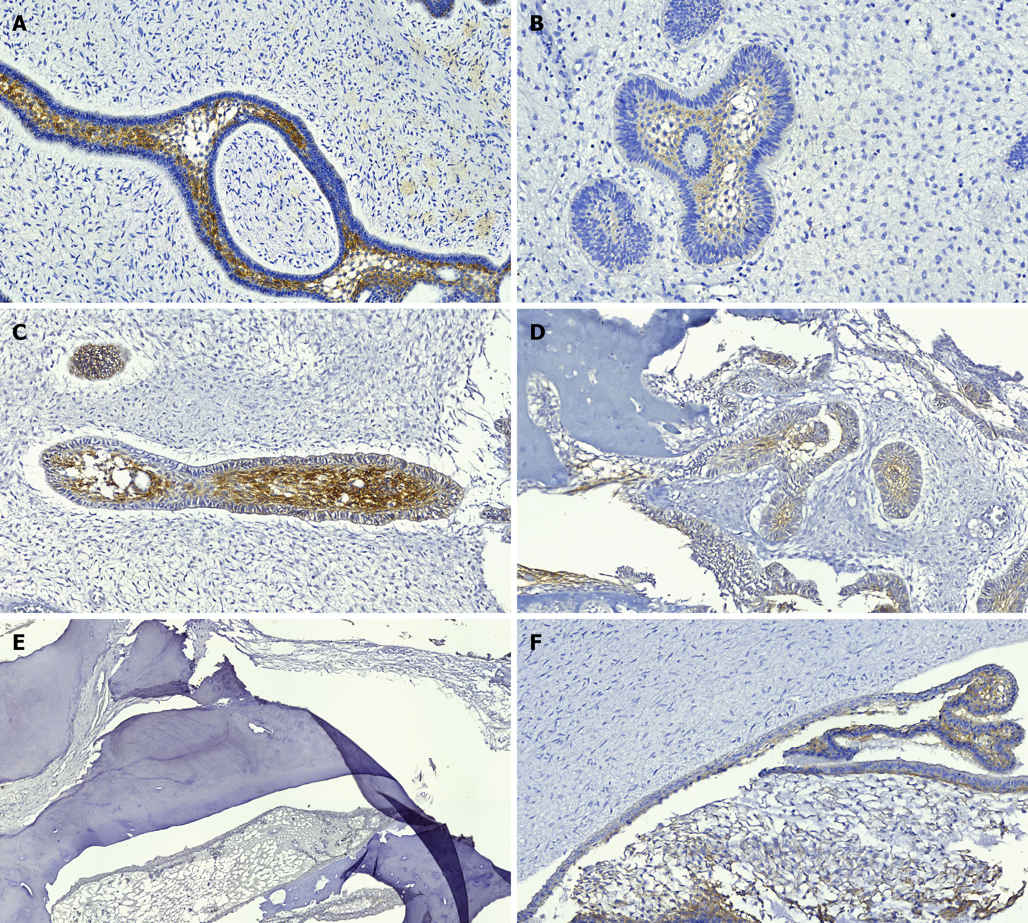
Figure 2 Immunoexpression of MOC-31 in ameloblastic fibrodentinoma, ameloblastic fibroma, ameloblastic fibro-odontoma, odontoma, and pericoronal cap.
A: Ameloblastic fibrodentinoma with positivity (++) in the stellate reticulum; B: Ameloblastic fibroma with positivity (+) in the stellate reticulum; C: Ameloblastic fibro-odontoma showing positivity (++) in stellate reticulum areas and ameloblasts; D: Ameloblastic fibro-odontoma where positivity is observed in epithelial islands continuous with the formation of mineralized structures; E: Odontoma, which was negative for protein immunoexpression; F: Pericoronal cap with positivity (+) in odontogenic epithelial tissues. Immunohistochemistry, MOC-31, A-F: × 20.
- Citation: Umpiérrez NG, Sicco E, Silveira F, Beovide AV, Bologna-Molina R. Differences of immunohistochemical profiling of MOC-31, caveolin-1, connexin-43, Ki-67 in ameloblastic fibroma, ameloblastic fibrodentinoma, ameloblastic fibro-odontoma, and odontomas. World J Exp Med 2026; 16(1): 115508
- URL: https://www.wjgnet.com/2220-315x/full/v16/i1/115508.htm
- DOI: https://dx.doi.org/10.5493/wjem.v16.i1.115508