
















©The Author(s) 2025.
World J Exp Med. Dec 20, 2025; 15(4): 114368
Published online Dec 20, 2025. doi: 10.5493/wjem.v15.i4.114368
Published online Dec 20, 2025. doi: 10.5493/wjem.v15.i4.114368
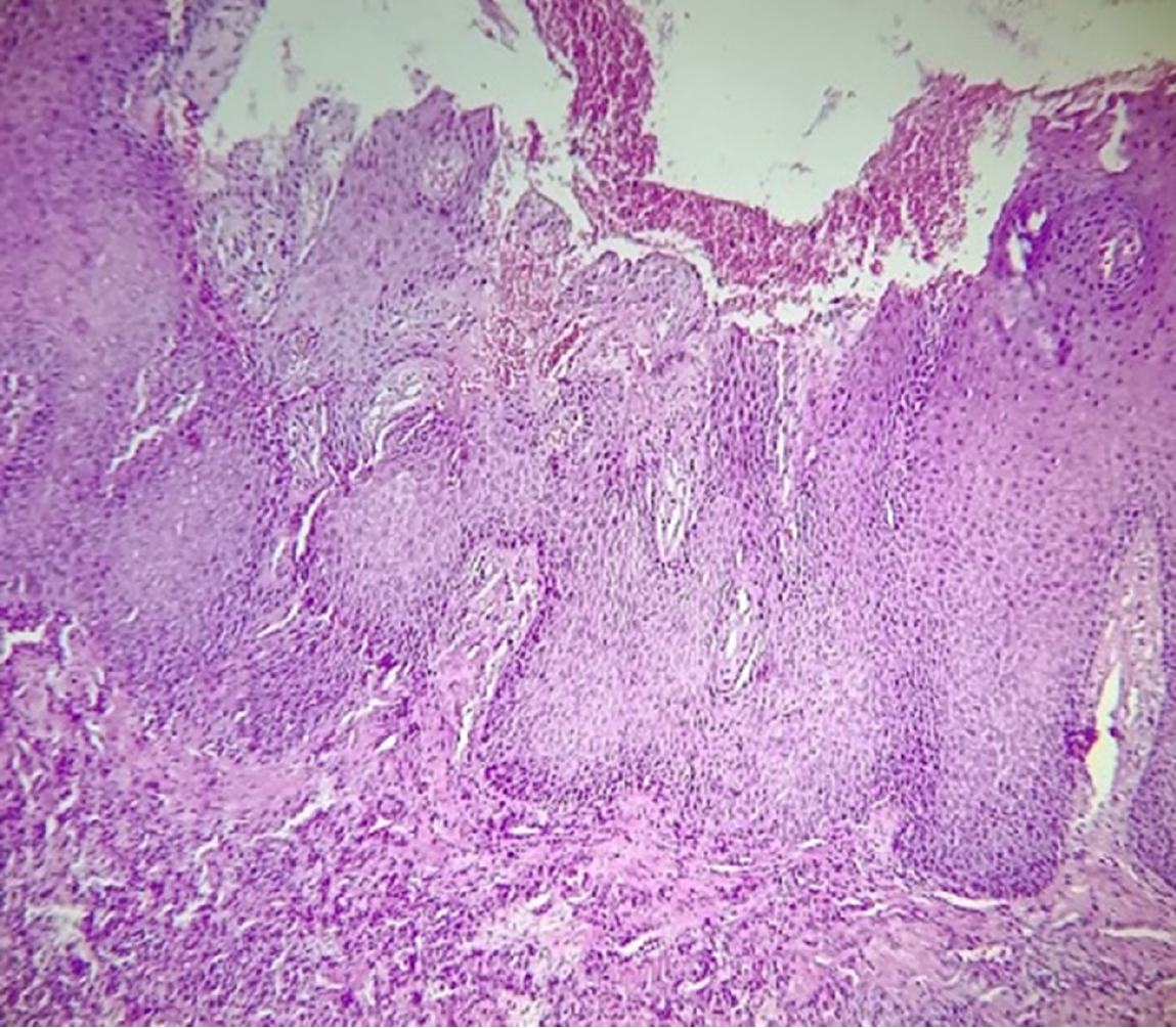
Figure 4 In multifocal epithelial hyperplasia samples, the stratified squamous epithelium presents pronounced hyperorthokeratosis, accompanied by acanthosis and the presence of koilocytes in the intermediate layers.
At the base of the epithelium, dense, well-vascularized connective tissue with chronic inflammatory infiltration is present, which is distributed abundantly and diffusely (100 ×).
- Citation: Ramos-Gregorio CO, Tremillo-Maldonado O, Silveira F, Schuch LF, Pereira-Prado V, Sicco E, Soto-Najera AC, GómezPalacio-Gastélum M, Isiordia-Espinoza M, Muñoz-Ibarra JJ, Toral-Rizo V, Bologna-Molina R. Multifocal epithelial hyperplasia: Clinical features, diagnosis and management challenges. World J Exp Med 2025; 15(4): 114368
- URL: https://www.wjgnet.com/2220-315x/full/v15/i4/114368.htm
- DOI: https://dx.doi.org/10.5493/wjem.v15.i4.114368