
















©The Author(s) 2025.
World J Exp Med. Dec 20, 2025; 15(4): 114368
Published online Dec 20, 2025. doi: 10.5493/wjem.v15.i4.114368
Published online Dec 20, 2025. doi: 10.5493/wjem.v15.i4.114368
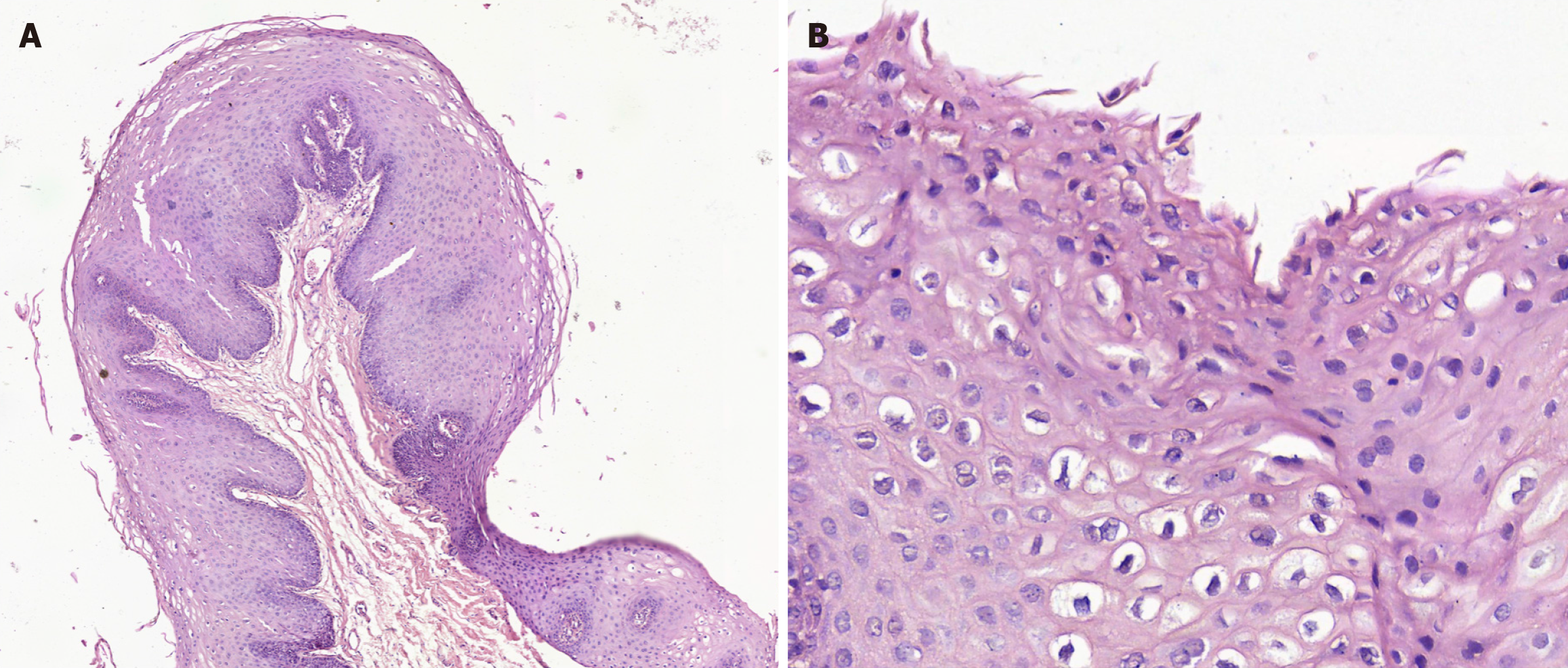
Figure 3 Histopatological image.
A: A lesion characterized by the presence of a stratified squamous epithelium parakeratinized with acanthosis and thick and elongated epithelial processes, which give a papillomatous appearance. In some areas, mitosoid and koilocytic figures are observed (50 ×). The connective tissue was well vascularized and free of injury; B: Mitosoid and koilocytic figures (200 ×).
- Citation: Ramos-Gregorio CO, Tremillo-Maldonado O, Silveira F, Schuch LF, Pereira-Prado V, Sicco E, Soto-Najera AC, GómezPalacio-Gastélum M, Isiordia-Espinoza M, Muñoz-Ibarra JJ, Toral-Rizo V, Bologna-Molina R. Multifocal epithelial hyperplasia: Clinical features, diagnosis and management challenges. World J Exp Med 2025; 15(4): 114368
- URL: https://www.wjgnet.com/2220-315x/full/v15/i4/114368.htm
- DOI: https://dx.doi.org/10.5493/wjem.v15.i4.114368