
















©The Author(s) 2025.
World J Exp Med. Dec 20, 2025; 15(4): 108187
Published online Dec 20, 2025. doi: 10.5493/wjem.v15.i4.108187
Published online Dec 20, 2025. doi: 10.5493/wjem.v15.i4.108187
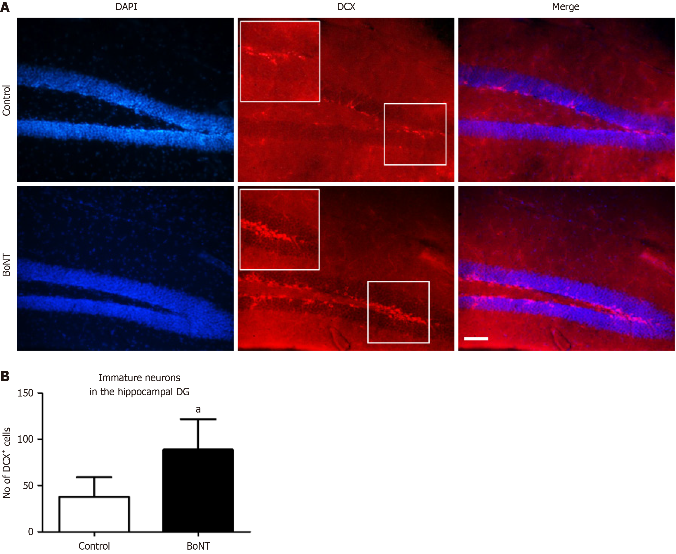
Figure 1 Immunohistochemical analysis of doublecortin-positive cells in the hippocampal dentate gyrus.
A: Representative image of doublecortin (DCX) immuno-positive cells in hippocampal dentate gyrus of control and botulinum neurotoxin (BoNT)-treated mice. The scale bar = 25 μm; B: The bar graph represents the number of DCX-positive cells. The number of DCX-positive cells in the BoNT was significantly increased when compared to the control group (aP value ≤ 0.01). DG: Dentate gyrus; BoNT: Botulinum neurotoxin; DCX: Doublecortin.
- Citation: Joseph JHM, Babu Deva Irakkam MP, Kandasamy M. Proneurogenic and microglial modulatory properties of botulinum neurotoxin in the hippocampus of aging experimental mice. World J Exp Med 2025; 15(4): 108187
- URL: https://www.wjgnet.com/2220-315x/full/v15/i4/108187.htm
- DOI: https://dx.doi.org/10.5493/wjem.v15.i4.108187