Copyright
©The Author(s) 2025.
World J Exp Med. Jun 20, 2025; 15(2): 100443
Published online Jun 20, 2025. doi: 10.5493/wjem.v15.i2.100443
Published online Jun 20, 2025. doi: 10.5493/wjem.v15.i2.100443
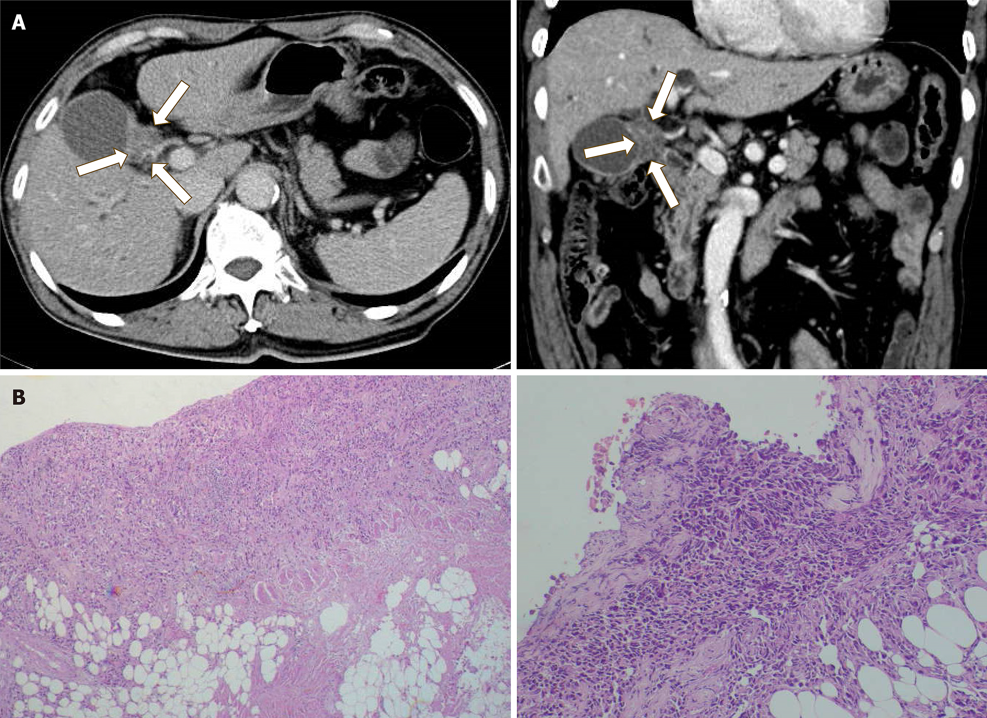
Figure 1 Computed tomography scan and histopathological findings of a patient with gall bladder cancer.
A: Computed tomography (CT) findings of the primary tumor. CT scan image of the transverse and coronal planes display gallbladder carcinoma (arrows); B: Hematoxylin and eosin staining of the metastatic peritoneal tumor of patients with gallbladder cancer. The tumor showed poorly differentiated growth with moderate stromal cells.
- Citation: Wang Q, Fan C, Tsujio G, Sakuma T, Maruo K, Yamamoto Y, Imanishi D, Kawabata K, Nishikubo H, Kanei S, Aoyama R, Kushiyama S, Ohira M, Yashiro M. Establishment and characterization of a new human gallbladder cancer cell line, OCUG-2. World J Exp Med 2025; 15(2): 100443
- URL: https://www.wjgnet.com/2220-315x/full/v15/i2/100443.htm
- DOI: https://dx.doi.org/10.5493/wjem.v15.i2.100443


