Copyright
©2013 Baishideng Publishing Group Co.
World J Exp Med. Nov 20, 2013; 3(4): 100-107
Published online Nov 20, 2013. doi: 10.5493/wjem.v3.i4.100
Published online Nov 20, 2013. doi: 10.5493/wjem.v3.i4.100
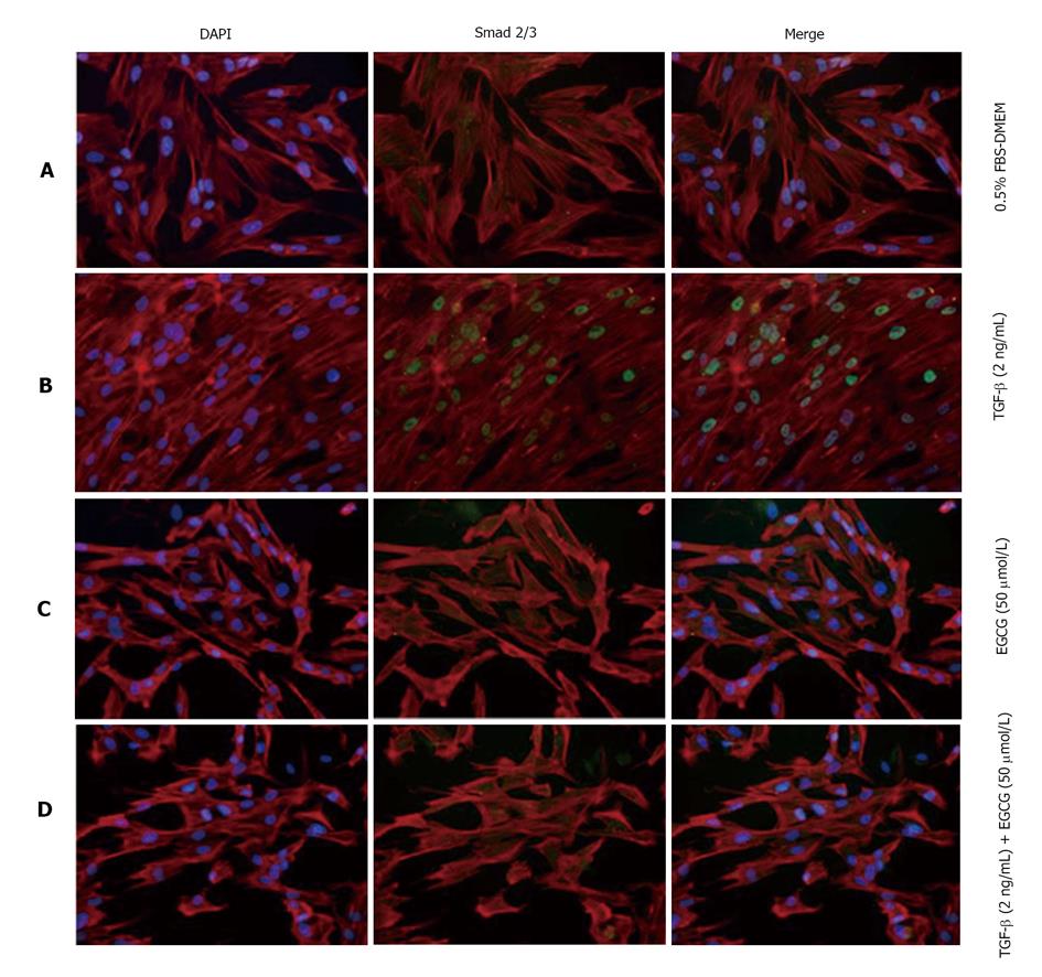
Figure 3 Effects of (-)-epigallocatechin-3-gallate on activation and localization of Smad2/3.
MRC-5 cells were treated with transforming growth factor-β (TGF-β) and/or (-)-epigallocatechin-3-gallate (EGCG). Cells were examined by confocal microscopy. Subcellular localization of Smad2/3 (green) and actin stress fibers (red) are shown. Nuclei were stained by DAPI (blue). A: Control; B: Treated with TGF-β; C: Treated with EGCG; D: Treated with TGF-β and EGCG. DAPI: 4',6-diamidino-2-phenylindole.
- Citation: Tabuchi M, Hayakawa S, Honda E, Ooshima K, Itoh T, Yoshida K, Park AM, Higashino H, Isemura M, Munakata H. Epigallocatechin-3-gallate suppresses transforming growth factor-beta signaling by interacting with the transforming growth factor-beta type II receptor. World J Exp Med 2013; 3(4): 100-107
- URL: https://www.wjgnet.com/2220-315X/full/v3/i4/100.htm
- DOI: https://dx.doi.org/10.5493/wjem.v3.i4.100


