Copyright
©The Author(s) 2025.
World J Exp Med. Sep 20, 2025; 15(3): 107248
Published online Sep 20, 2025. doi: 10.5493/wjem.v15.i3.107248
Published online Sep 20, 2025. doi: 10.5493/wjem.v15.i3.107248
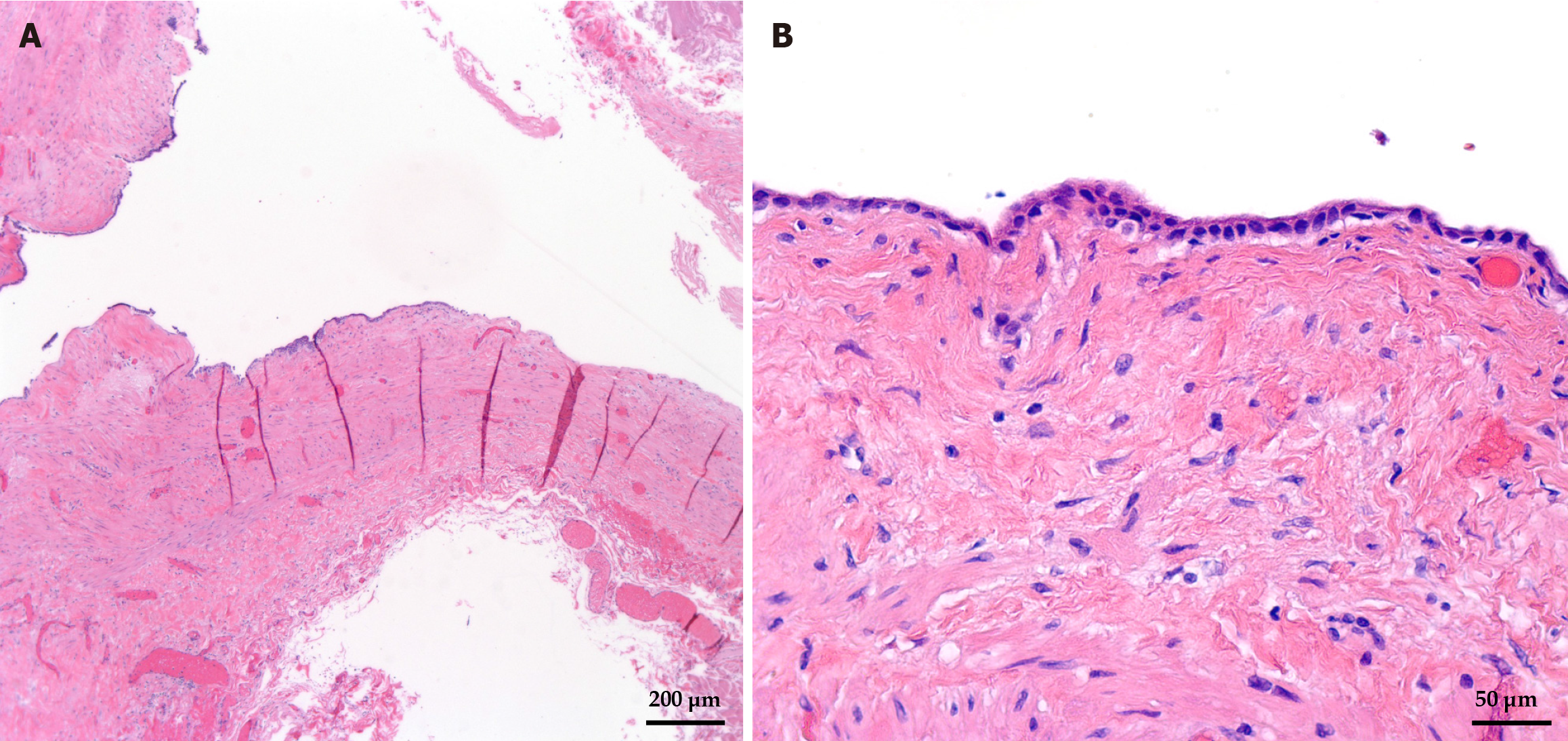
Figure 4 Microscopic images of the gallbladder mucocele.
A: Hematoxylin and eosin-stained sections of the gallbladder mucocele wall; B: High-power hematoxylin and eosin-stained section of the mucocele wall lining showing atrophic cuboidal epithelium.
- Citation: Thiravialingam A, Sriganeshan K, Bahmad HF, Polit F, Ahmed A, Joshi D, Poppiti R. Incidental gallbladder mucocele mimicking acute cholecystitis: A case report and review of literature. World J Exp Med 2025; 15(3): 107248
- URL: https://www.wjgnet.com/2220-315X/full/v15/i3/107248.htm
- DOI: https://dx.doi.org/10.5493/wjem.v15.i3.107248


