
















©The Author(s) 2025.
World J Exp Med. Sep 20, 2025; 15(3): 107248
Published online Sep 20, 2025. doi: 10.5493/wjem.v15.i3.107248
Published online Sep 20, 2025. doi: 10.5493/wjem.v15.i3.107248
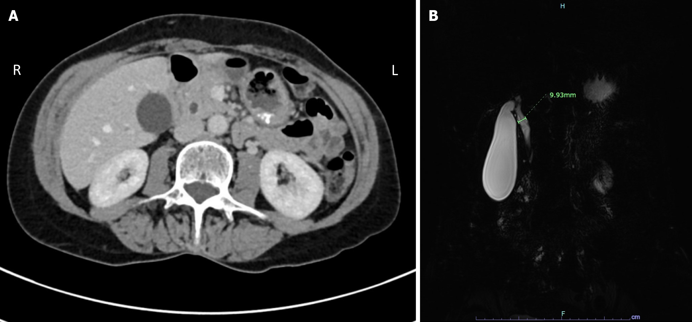
Figure 1 Computed tomography scan and magnetic resonance imaging without contrast image studies.
A: Computed tomography scan of the abdomen and pelvis demonstrating dilatation of the central biliary ducts tapering smoothly towards the ampulla without a discrete obstructing lesion. The gallbladder is distended without wall thickening or acute inflammation; B: Magnetic resonance imaging without contrast shows a hydropic gallbladder without wall thickening or pericholecystic inflammatory changes. The common bile duct is dilated measuring up to 10 mm in the proximal third of the duct with smooth gradual tapering to normal caliber towards the ampullary region. No choledocholithiasis has been identified.
- Citation: Thiravialingam A, Sriganeshan K, Bahmad HF, Polit F, Ahmed A, Joshi D, Poppiti R. Incidental gallbladder mucocele mimicking acute cholecystitis: A case report and review of literature. World J Exp Med 2025; 15(3): 107248
- URL: https://www.wjgnet.com/2220-315X/full/v15/i3/107248.htm
- DOI: https://dx.doi.org/10.5493/wjem.v15.i3.107248