Copyright
©The Author(s) 2016.
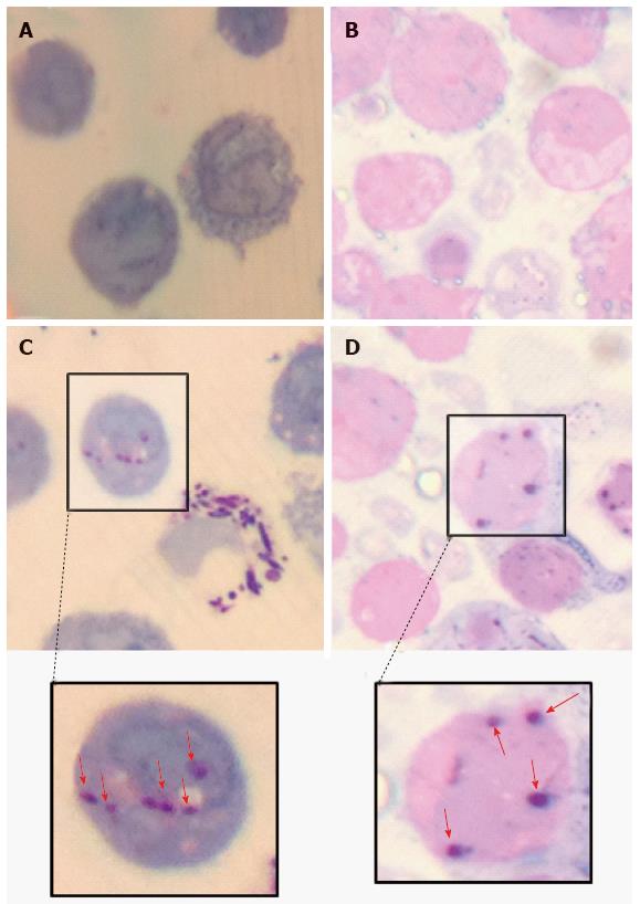
Figure 8 Light microscopic evidence of the intracellular presence of Mycobacterium marinum in human mast cell line.
Gram-Twort stained, 0.5 µm thin cross sections of Mono Mac 6 cells [A: uninfected; C: infected with Mycobacterium marinum (M. marinum) at MOI 10 for 24 h] and human mast cell line (HMC-1) cells (B: Uninfected; D: Infected HMC-1 cells with M. marinum at MOI 10 for 24 h). The red arrows in insets C and D indicate internalised M. marinum (Olympus CH2, × 100 oil immersion, captured using iPhone 4).
- Citation: Siad S, Byrne S, Mukamolova G, Stover C. Intracellular localisation of Mycobacterium marinum in mast cells. World J Immunol 2016; 6(1): 83-95
- URL: https://www.wjgnet.com/2219-2824/full/v6/i1/83.htm
- DOI: https://dx.doi.org/10.5411/wji.v6.i1.83


