
















©The Author(s) 2026.
World J Clin Urol. Feb 12, 2026; 15(1): 114046
Published online Feb 12, 2026. doi: 10.5410/wjcu.v15.i1.114046
Published online Feb 12, 2026. doi: 10.5410/wjcu.v15.i1.114046
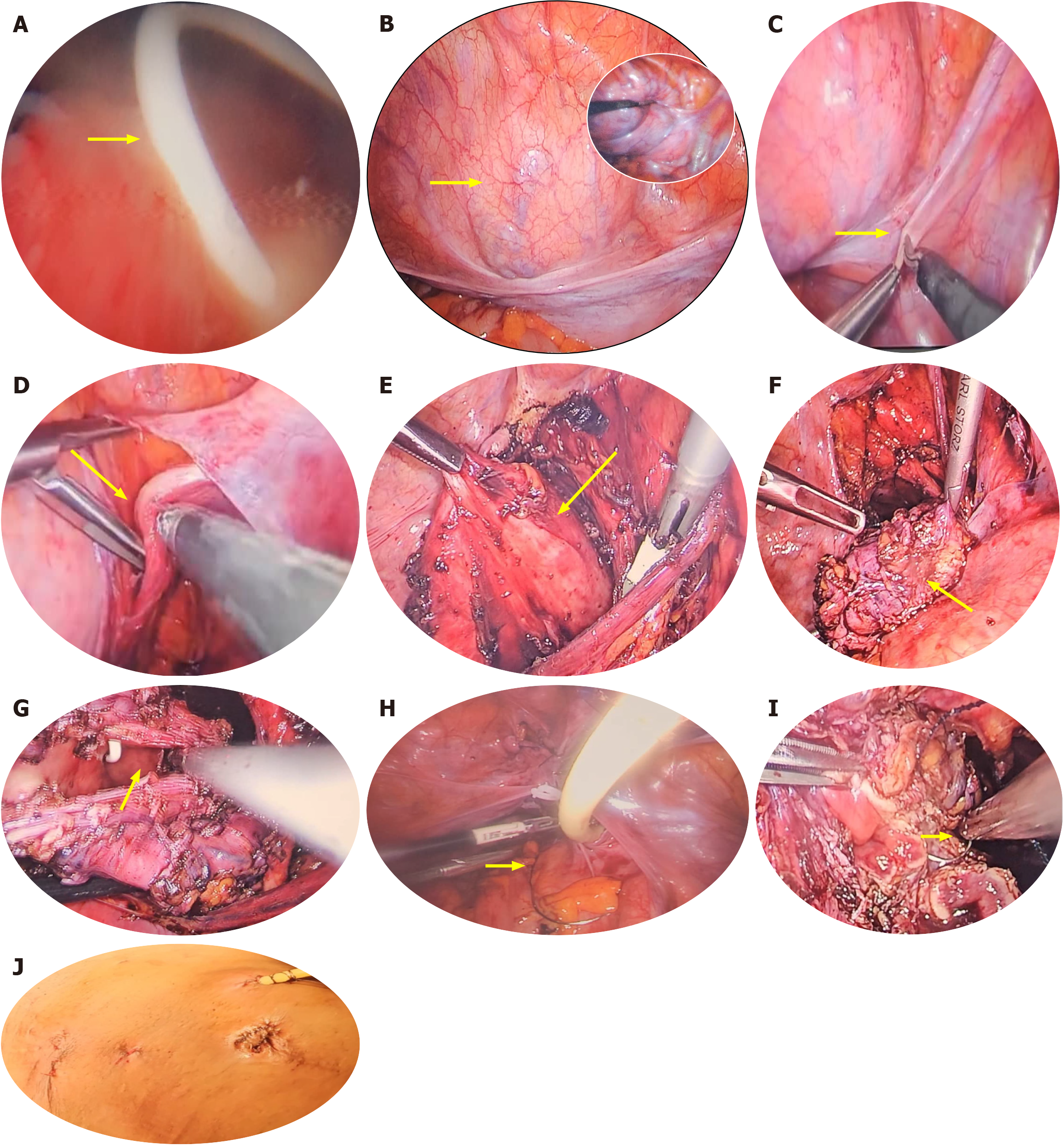
Figure 10 Laparoscopic bladder diverticulectomy demonstrating various stages of laparoscopic extravesical bladder diverticulectomy.
A: Initial cystoscopy and stenting of the adjacent ureter (arrow); B: Combined endoscopic and laparoscopic localization of the bladder diverticulum (arrow) with the inset; C: Identification of anatomical landmarks and initiation of laparoscopic dissection (arrow); D: Dissection of the vas deferens (arrow); E: Dissection of the diverticulum sac (arrow); F: Completion of diverticulum sac dissection (arrow); G: Assessment of the diverticular neck and the adjacent ureter before closure (arrow); H and I: Closure of the diverticular neck (arrow); J: Immediate postoperative view of the abdomen (arrow).
- Citation: Khalid A, Obadele OG, Alabi TO, Nedjim SA, Abdulwahab-Ahmed A, Mungadi IA. Practical approach to the review of bladder diverticulum and its management. World J Clin Urol 2026; 15(1): 114046
- URL: https://www.wjgnet.com/2219-2816/full/v15/i1/114046.htm
- DOI: https://dx.doi.org/10.5410/wjcu.v15.i1.114046