
















©The Author(s) 2026.
World J Clin Urol. Feb 12, 2026; 15(1): 114046
Published online Feb 12, 2026. doi: 10.5410/wjcu.v15.i1.114046
Published online Feb 12, 2026. doi: 10.5410/wjcu.v15.i1.114046
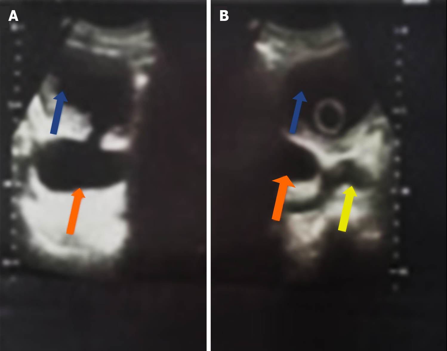
Figure 2 Transabdominal ultrasound scan.
A: Displays a paraureteral diverticulum (orange arrows), bladder (blue arrows), and the connection by the diverticular neck; B: The same patient demonstrates how the paraureteral diverticulum protrudes under the bladder neck during voiding, thus causing urinary retention. The paraureteral diverticulum (orange arrows), bladder with an indwelling catheter (blue arrows), and the protruded paraureteral diverticulum under the bladder neck (yellow arrow) are all noted.
- Citation: Khalid A, Obadele OG, Alabi TO, Nedjim SA, Abdulwahab-Ahmed A, Mungadi IA. Practical approach to the review of bladder diverticulum and its management. World J Clin Urol 2026; 15(1): 114046
- URL: https://www.wjgnet.com/2219-2816/full/v15/i1/114046.htm
- DOI: https://dx.doi.org/10.5410/wjcu.v15.i1.114046