
















©The Author(s) 2025.
World J Clin Urol. Mar 18, 2025; 14(1): 104791
Published online Mar 18, 2025. doi: 10.5410/wjcu.v14.i1.104791
Published online Mar 18, 2025. doi: 10.5410/wjcu.v14.i1.104791
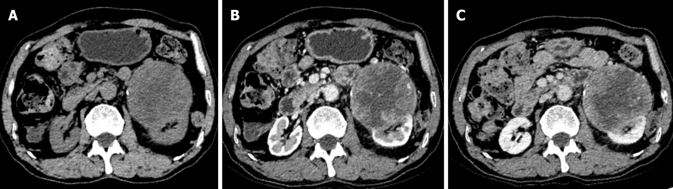
Figure 1 The preoperative computed tomography images of the patient.
A: The plain scan of computed tomography examination shows a cystic- solid mass shadow with uneven density; B: After enhanced scanning, it shows heterogeneous enhancement; C: The solid part in the venous phase shows a "fast in and slow out" performance.
- Citation: Deng CH, Zhou Y, Chen J, He GF, Fu QS, Li JM, Wang G, Hu XD. Diagnostic and therapeutic considerations for primary penal synovial sarcoma with specific MDM2 gene amplification: A case report. World J Clin Urol 2025; 14(1): 104791
- URL: https://www.wjgnet.com/2219-2816/full/v14/i1/104791.htm
- DOI: https://dx.doi.org/10.5410/wjcu.v14.i1.104791