Copyright
©The Author(s) 2021.
World J Clin Urol. Apr 24, 2021; 10(1): 1-6
Published online Apr 24, 2021. doi: 10.5410/wjcu.v10.i1.1
Published online Apr 24, 2021. doi: 10.5410/wjcu.v10.i1.1
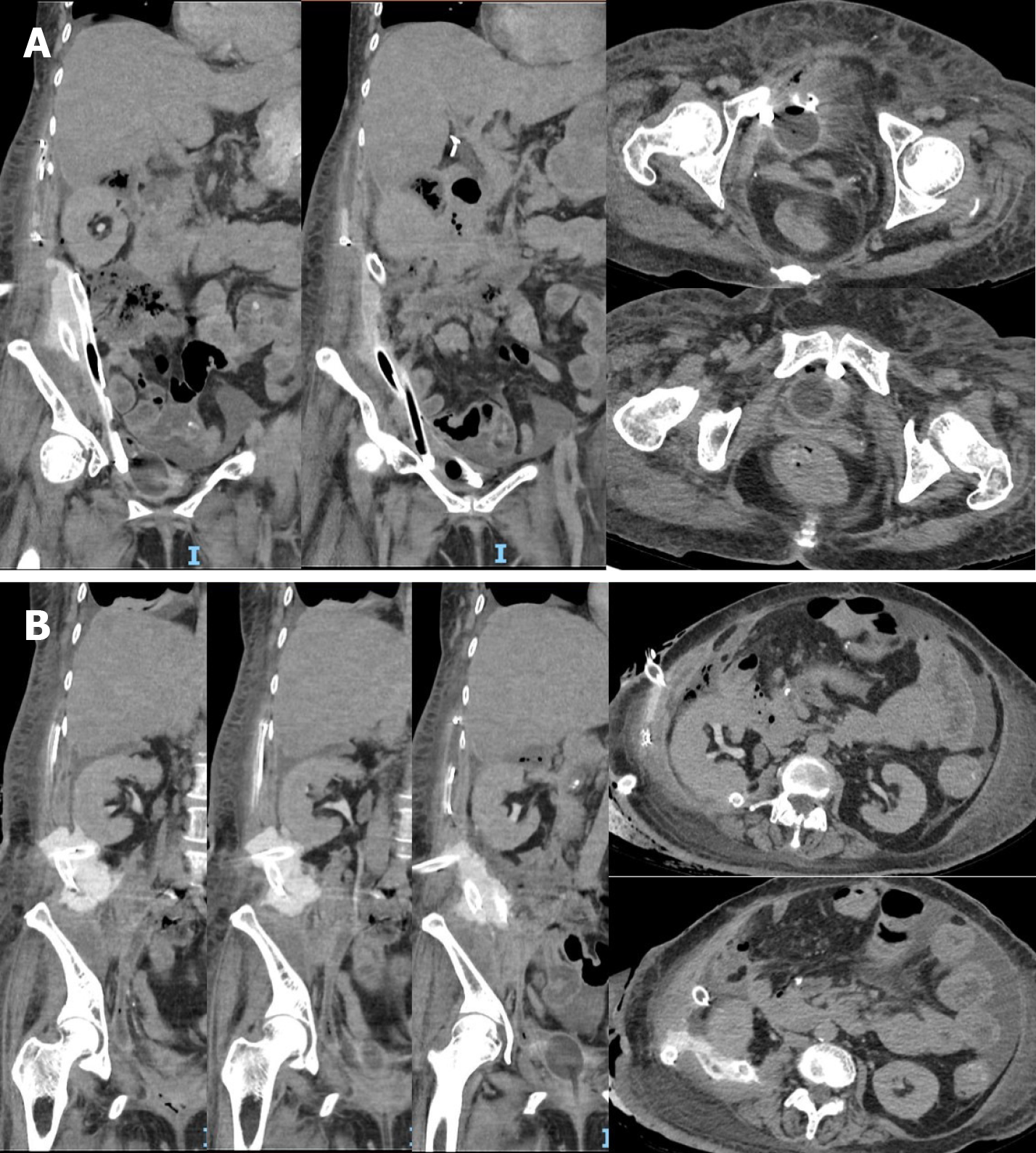
Figure 1 Coronal and axial views of computed tomography urography.
A: Drains through right upper quadrant and flank directed inferiorly into the pelvis; B: The perinephric urinoma and right flank drain directed posterior to the right kidney.
- Citation: Kumaran A, Yeung PM, Tiwari R. Perinephric urinoma, an unusual upper tract presentation of a lower tract injury following retroperitoneoscopy: A case report. World J Clin Urol 2021; 10(1): 1-6
- URL: https://www.wjgnet.com/2219-2816/full/v10/i1/1.htm
- DOI: https://dx.doi.org/10.5410/wjcu.v10.i1.1


