Copyright
©The Author(s) 2019.
World J Stomatol. Dec 18, 2019; 7(3): 28-38
Published online Dec 18, 2019. doi: 10.5321/wjs.v7.i3.28
Published online Dec 18, 2019. doi: 10.5321/wjs.v7.i3.28
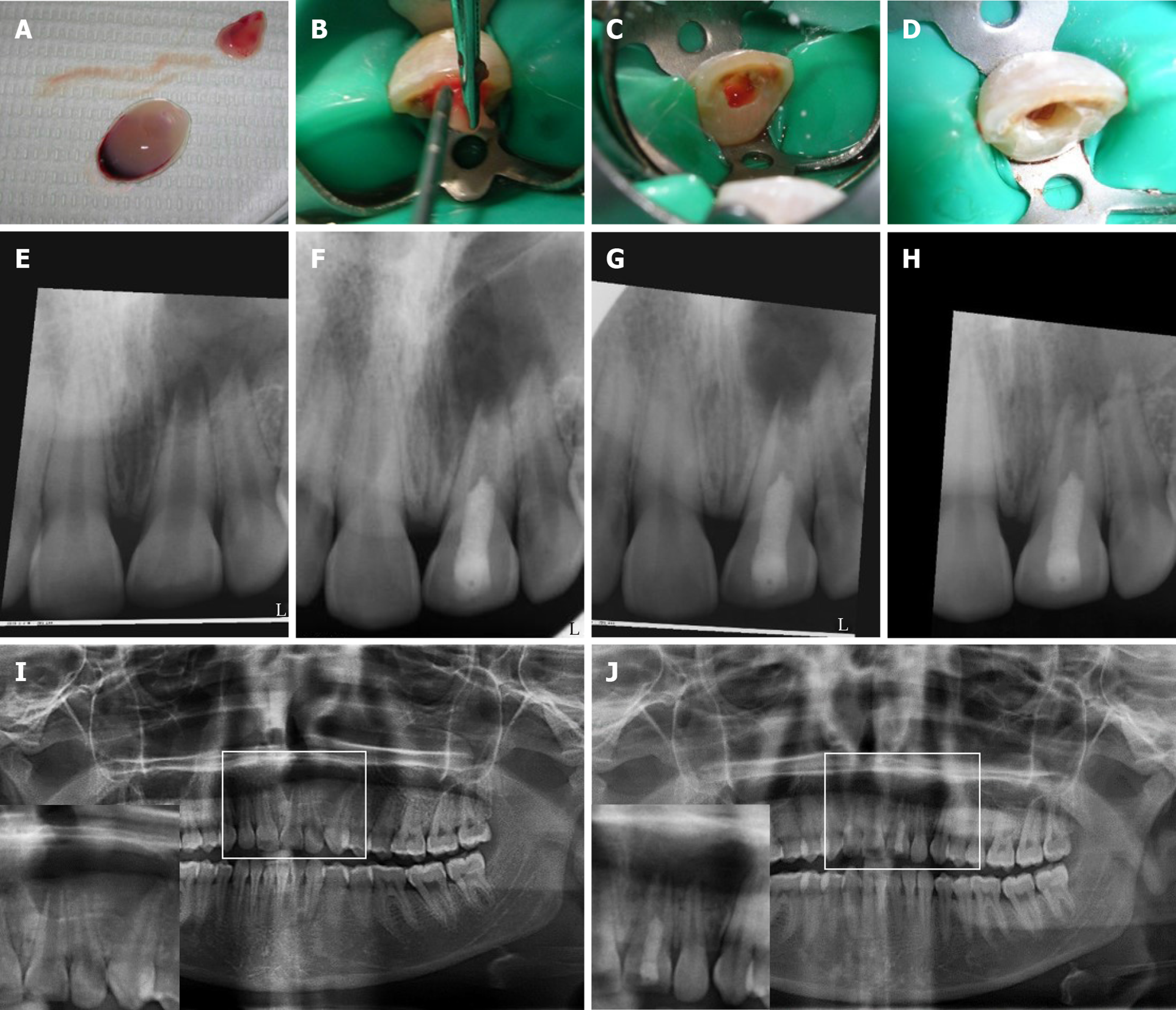
Figure 2 Revascularization procedure using platelet-rich fibrin and follow-up digital periapical radiographs and orthopantograms for case #1 (tooth #9).
A-D: Prepared platelet-rich fibrin (PRF) clots in A with insert showing squeezing of clots in a metallic mesh to release supernatant (extract); B: Packing of PRF clot into the root canal with platelet-plug facing apical tissues using sterile cotton pliers and endodontic pluggers; and removal of excess extract and cleaning of pulp chamber in C and D, respectively; E, F: Periapical radiographs of the case showing in E pre-operative radiograph; F: after 1 mo; G: After 3 mo and; H: After 9 mo; I, J: Show pre- and 9 mo post-revascularization panoramic radiographs using the same device, respectively. Inserts represent magnifications of the selected regions of interest shown in I and J, respectively. Follow-up radiographs show progress in healing of the periapical lesion in terms of both size and density. The lesion originally extended from the maxillary left central incisor to the mesial root surface of the left canine. Teeth were asymptomatic. The case was unfortunately lost to follow-up after 9 mo. All digital periapical radiographs were aligned using the Turboreg plugin- image J software (http://bigwww.epfl.ch/thevenaz/turboreg/).
- Citation: Eltawila AM, El Backly R. Autologous platelet-rich-fibrin-induced revascularization sequelae: Two case reports. World J Stomatol 2019; 7(3): 28-38
- URL: https://www.wjgnet.com/2218-6263/full/v7/i3/28.htm
- DOI: https://dx.doi.org/10.5321/wjs.v7.i3.28


