Copyright
©2013 Baishideng.
World J Otorhinolaryngol. Aug 28, 2013; 3(3): 89-99
Published online Aug 28, 2013. doi: 10.5319/wjo.v3.i3.89
Published online Aug 28, 2013. doi: 10.5319/wjo.v3.i3.89
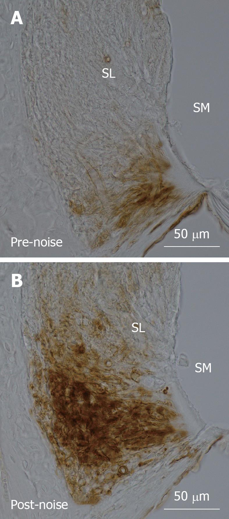
Figure 1 Intercellular adhesion molecule-1 immunolabelling in the spiral ligament of the cochlear basal turn in C57BL/6 mice.
A: In the non-noise exposed cochlea, intercellular adhesion molecule-1 (ICAM-1) was expressed by type IV fibrocytes and vascular endothelial cells in the lowest region of the spiral ligament; B: Mice exposed to traumatic noise (100 dB SPL, 8-16 kHz) for 24 h showed increased expression of ICAM-1, peaking at 24 h following acoustic trauma. ICAM-1 immunolabelling became more intense and expanded to cover a much greater area in the inferior region of the spiral ligament. ICAM-1 immunoexpression was determined by immunoperoxidase histochemistry and photomicrographs of mid-modiolar cochlear sections were taken with a digital light microscope (Nikon Eclipse 80i) at 40 × magnification. SL: Spiral ligament; SM: Scala media.
- Citation: Tan WJ, Thorne PR, Vlajkovic SM. Noise-induced cochlear inflammation. World J Otorhinolaryngol 2013; 3(3): 89-99
- URL: https://www.wjgnet.com/2218-6247/full/v3/i3/89.htm
- DOI: https://dx.doi.org/10.5319/wjo.v3.i3.89


