Copyright
©2013 Baishideng.
World J Otorhinolaryngol. Feb 28, 2013; 3(1): 1-15
Published online Feb 28, 2013. doi: 10.5319/wjo.v3.i1.1
Published online Feb 28, 2013. doi: 10.5319/wjo.v3.i1.1
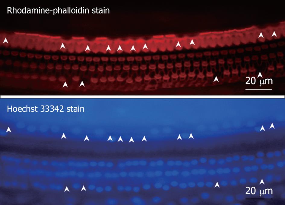
Figure 8 Fluorescence microscopic findings of the organ of Corti 7 d after ischemic insult.
The specimen was stained with rhodamine-phalloidin and Hoechst 33342. Three rows of outer hair cells (OHC) and a single row of inner hair cells (IHC) could be observed, and the cell loss was more severe in IHC than OHC. Arrowheads indicate loss of hair cells.
- Citation: Gyo K. Experimental study of transient cochlear ischemia as a cause of sudden deafness. World J Otorhinolaryngol 2013; 3(1): 1-15
- URL: https://www.wjgnet.com/2218-6247/full/v3/i1/1.htm
- DOI: https://dx.doi.org/10.5319/wjo.v3.i1.1


