Copyright
©2014 Baishideng Publishing Group Inc.
World J Ophthalmol. Aug 12, 2014; 4(3): 75-81
Published online Aug 12, 2014. doi: 10.5318/wjo.v4.i3.75
Published online Aug 12, 2014. doi: 10.5318/wjo.v4.i3.75
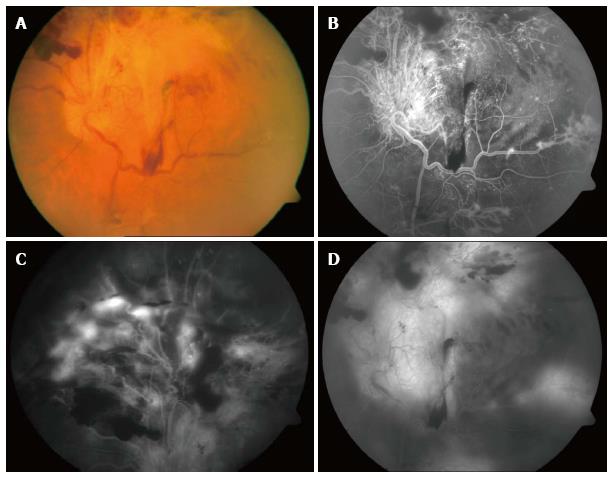
Figure 1 Preoperative images of the retina showing diabetic retinal detachment involving the posterior pole (A), the superior retina (B), and the region near the arcade (C), as well as subhyaloid and preretinal hemorrhage in the equator (D).
- Citation: Ferreira MA, Ferreira REA, Silva NS. Preoperative intravitreal bevacizumab and silicone oil tamponade for vitrectomy in diabetic retinopathy. World J Ophthalmol 2014; 4(3): 75-81
- URL: https://www.wjgnet.com/2218-6239/full/v4/i3/75.htm
- DOI: https://dx.doi.org/10.5318/wjo.v4.i3.75


