Copyright
©The Author(s) 2015.
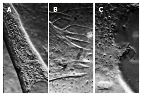
Figure 8 Differential interference contrast images of three different cell types (mouse myotube, A; rat astrocyte, B, human embryonic kidney cell, C) showing the variability of membrane structure and how patch clamp recordings are expected to be variable.
The structures above change rapidly over time (this is a frame from a movie, Courtesy Suchyna T).
- Citation: Sachs F. Mechanical transduction by ion channels: A cautionary tale. World J Neurol 2015; 5(3): 74-87
- URL: https://www.wjgnet.com/2218-6212/full/v5/i3/74.htm
- DOI: https://dx.doi.org/10.5316/wjn.v5.i3.74


