
















©The Author(s) 2015.
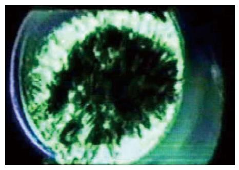
Figure 7 A frame from tomographic reconstruction of a patch of a Xenopus oocyte using high voltage electron microscope tomography[1].
The image shows cytoskeleton spanning the pipette and the bilayer is attached to the upper side but is not visible in this reconstruction due to its low density.
- Citation: Sachs F. Mechanical transduction by ion channels: A cautionary tale. World J Neurol 2015; 5(3): 74-87
- URL: https://www.wjgnet.com/2218-6212/full/v5/i3/74.htm
- DOI: https://dx.doi.org/10.5316/wjn.v5.i3.74