
















©The Author(s) 2020.
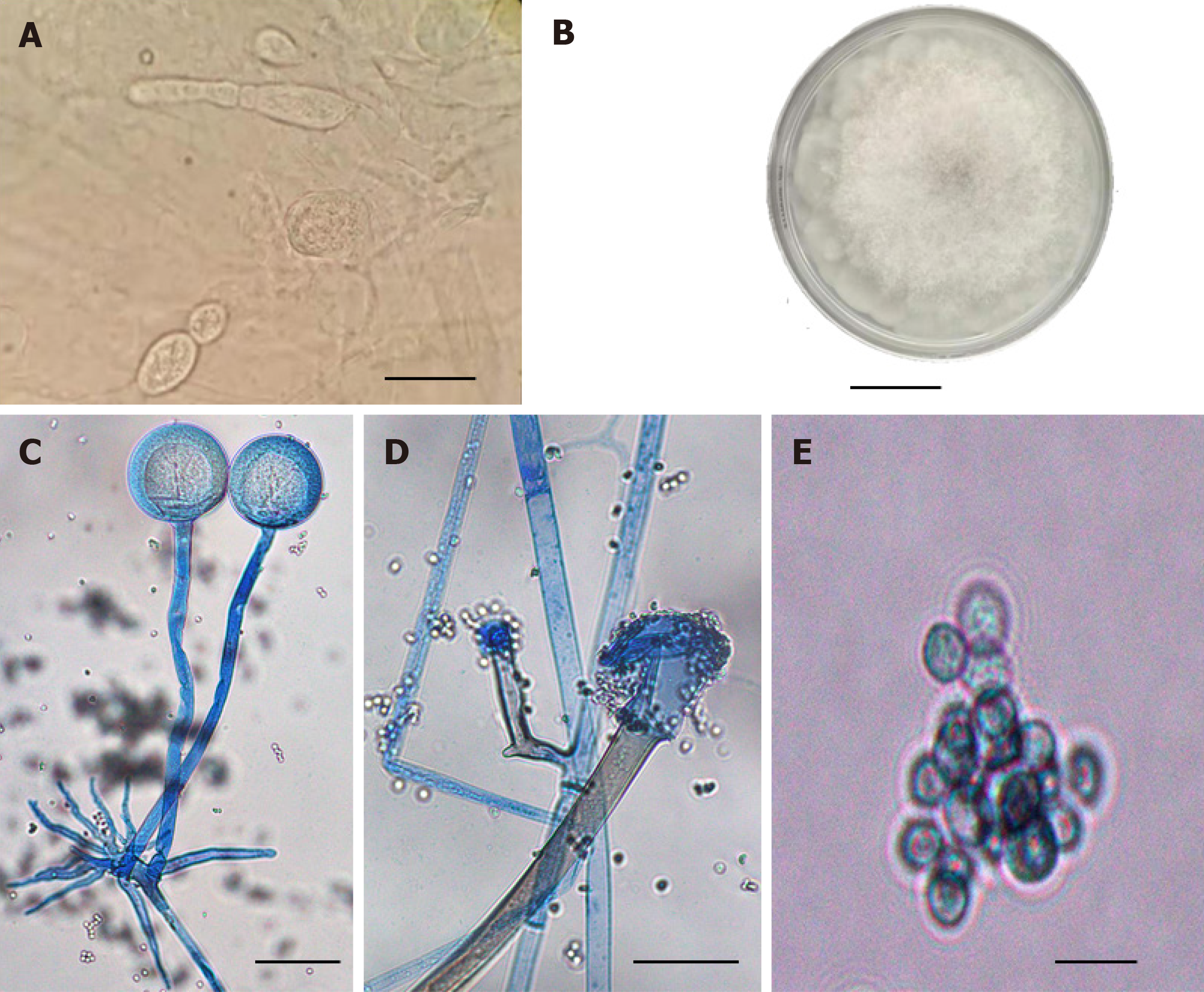
Figure 2 Pathological findings.
A: The patient's secretion was cultured at 33 °C for 3 d, and the morphology of fungal colony was observed; B: Typical sporangium and junction structure can be seen by direct microscopic examination of the patient's secretion; C: Typical spherical sporangium and developed rhizoid and sporophyte can be found by slide culture and lactophenol cotton blue staining; D: The shape of the sporangium after spores are released; E: Spore morphology of pathogenic fungi (regular round spore, with the range of diameter from 4.7 μm to 5.0 μm). All the scale bars represent 10 μm.
- Citation: Feng YH, Guo WW, Wang YR, Shi WX, Liu C, Li DM, Qiu Y, Shi DM. Rhinocerebral mucormycosis caused by Rhizopus oryzae in a patient with acute myeloid leukemia: A case report. World J Dermatol 2020; 8(1): 1-9
- URL: https://www.wjgnet.com/2218-6190/full/v8/i1/1.htm
- DOI: https://dx.doi.org/10.5314/wjd.v8.i1.1