Copyright
©The Author(s) 2016.
World J Dermatol. Nov 2, 2016; 5(4): 136-143
Published online Nov 2, 2016. doi: 10.5314/wjd.v5.i4.136
Published online Nov 2, 2016. doi: 10.5314/wjd.v5.i4.136
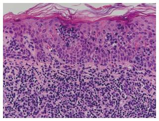
Figure 6 Skin specimen from patient 6 shows typical histopathologic features of mycosis fungoides.
Nests of medium to large sized neoplastic lymphocytes with pleomorphic and cerebriform nuclei are observed within the epidermis (Pautrier microabscesses) and adjacent superficial dermis (H and E, × 400).
- Citation: Vonderheid EC, Kadin ME, Telang GH. Papular mycosis fungoides: Six new cases and association with chronic lymphocytic leukemia. World J Dermatol 2016; 5(4): 136-143
- URL: https://www.wjgnet.com/2218-6190/full/v5/i4/136.htm
- DOI: https://dx.doi.org/10.5314/wjd.v5.i4.136


