
















©The Author(s) 2016.
World J Dermatol. Nov 2, 2016; 5(4): 136-143
Published online Nov 2, 2016. doi: 10.5314/wjd.v5.i4.136
Published online Nov 2, 2016. doi: 10.5314/wjd.v5.i4.136
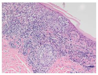
Figure 4 Skin specimen from patient 5 shows a moderately dense lichenoid infiltrate, wiry bundles of collagen in a thickened papillary dermis, and follicular mucinosis.
Numerous atypical lymphocytes, some with large irregular nuclei, are located within the epidermis, both as solitary units and in aggregates, and dermal infiltrate (H and E, × 400).
- Citation: Vonderheid EC, Kadin ME, Telang GH. Papular mycosis fungoides: Six new cases and association with chronic lymphocytic leukemia. World J Dermatol 2016; 5(4): 136-143
- URL: https://www.wjgnet.com/2218-6190/full/v5/i4/136.htm
- DOI: https://dx.doi.org/10.5314/wjd.v5.i4.136