Copyright
©The Author(s) 2016.
World J Dermatol. Nov 2, 2016; 5(4): 136-143
Published online Nov 2, 2016. doi: 10.5314/wjd.v5.i4.136
Published online Nov 2, 2016. doi: 10.5314/wjd.v5.i4.136
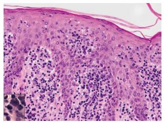
Figure 2 Skin specimen from patient 4 shows an acanthotic epidermis that contains atypical lymphocytes with hyperchromatic irregular nuclei (insert).
The dermis has a superficial infiltrate composed of normal and atypical lymphocytes (H and E, × 400).
- Citation: Vonderheid EC, Kadin ME, Telang GH. Papular mycosis fungoides: Six new cases and association with chronic lymphocytic leukemia. World J Dermatol 2016; 5(4): 136-143
- URL: https://www.wjgnet.com/2218-6190/full/v5/i4/136.htm
- DOI: https://dx.doi.org/10.5314/wjd.v5.i4.136


