Copyright
©The Author(s) 2015.
World J Dermatol. Nov 2, 2015; 4(4): 135-144
Published online Nov 2, 2015. doi: 10.5314/wjd.v4.i4.135
Published online Nov 2, 2015. doi: 10.5314/wjd.v4.i4.135
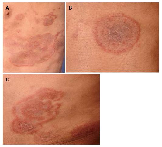
Figure 1 Mycosis fungoides presenting with annular lesions.
Clinical photographs of an 80-year-old male patient (case 1) with slowly progressive annular, cockadiform and arcuate lesions resembling tinea corporis (A), erythema exsudativum multiforme (B), and granuloma annulare (C), respectively.
- Citation: Wobser M, Geissinger E, Rosenwald A, Goebeler M. Mycosis fungoides: A mimicker of benign dermatoses. World J Dermatol 2015; 4(4): 135-144
- URL: https://www.wjgnet.com/2218-6190/full/v4/i4/135.htm
- DOI: https://dx.doi.org/10.5314/wjd.v4.i4.135


