Copyright
©The Author(s) 2015.
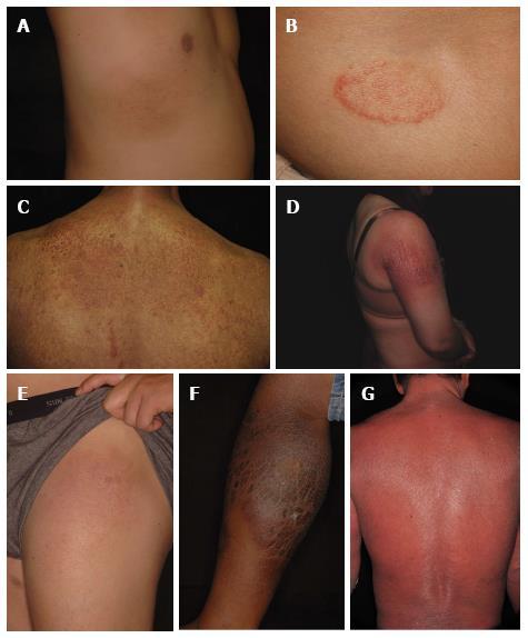
Figure 1 Clinical images showing patch stage (A), plaque stage (B-E), tumor stage (F), and erythrodermic (G) mycosis fungoides.
- Citation: Hu SCS. Mycosis fungoides and Sézary syndrome: Role of chemokines and chemokine receptors. World J Dermatol 2015; 4(2): 69-79
- URL: https://www.wjgnet.com/2218-6190/full/v4/i2/69.htm
- DOI: https://dx.doi.org/10.5314/wjd.v4.i2.69


