Copyright
©The Author(s) 2015.
World J Anesthesiol. Jul 27, 2015; 4(2): 30-38
Published online Jul 27, 2015. doi: 10.5313/wja.v4.i2.30
Published online Jul 27, 2015. doi: 10.5313/wja.v4.i2.30
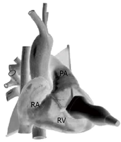
Figure 2 Parasternal right ventricular inflow outflow view, anterior projection.
Schematic diagram demonstrating transthoracic echocardiogram probe position and alignment of scanning plane. Reproduced in part with permission from Toronto General Hospital, Perioperative Interactive Education Virtual TTE (http://pie.med.utoronto.ca/TTE). TTE: Transthoracic echocardiogram; RA: Right atrium; RV: Right ventricle; PA: Pulmonary artery.
- Citation: Tan CO, Weinberg L, Story DA, McNicol L. Transthoracic echocardiography assists appropriate pulmonary artery catheter placement: An observational study. World J Anesthesiol 2015; 4(2): 30-38
- URL: https://www.wjgnet.com/2218-6182/full/v4/i2/30.htm
- DOI: https://dx.doi.org/10.5313/wja.v4.i2.30


