Copyright
©The Author(s) 2017.
World J Clin Oncol. Aug 10, 2017; 8(4): 305-319
Published online Aug 10, 2017. doi: 10.5306/wjco.v8.i4.305
Published online Aug 10, 2017. doi: 10.5306/wjco.v8.i4.305
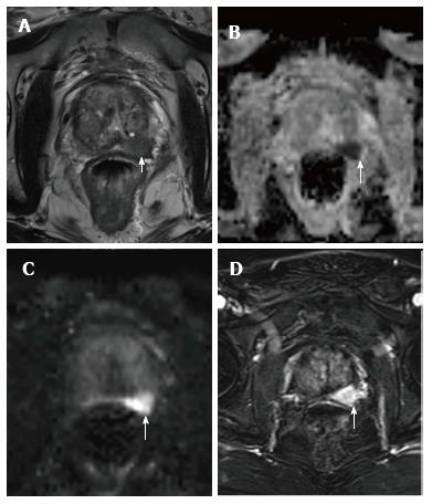
Figure 5 An 82-year-old patient with PIRADS 5 lesion in the basal left peripheral zone showing hypointensity (white arrows) in T2-weighted image (A), restriction of diffusion in apparent diffusion coefficient map (B) and diffusion-weighted magnetic resonance image (C), DCE shows significant early enhancement of the suspicious area (D).
MRI-guided transrectal ultrasound biopsy confirmed a clinically significant prostatic carcinoma (Gleason 5 + 4). DCE: Dynamic contrast enhancement; MRI: Magnetic resonance imaging.
- Citation: Couñago F, Sancho G, Catalá V, Hernández D, Recio M, Montemuiño S, Hernández JA, Maldonado A, del Cerro E. Magnetic resonance imaging for prostate cancer before radical and salvage radiotherapy: What radiation oncologists need to know. World J Clin Oncol 2017; 8(4): 305-319
- URL: https://www.wjgnet.com/2218-4333/full/v8/i4/305.htm
- DOI: https://dx.doi.org/10.5306/wjco.v8.i4.305


