Copyright
©The Author(s) 2017.
World J Clin Oncol. Aug 10, 2017; 8(4): 305-319
Published online Aug 10, 2017. doi: 10.5306/wjco.v8.i4.305
Published online Aug 10, 2017. doi: 10.5306/wjco.v8.i4.305
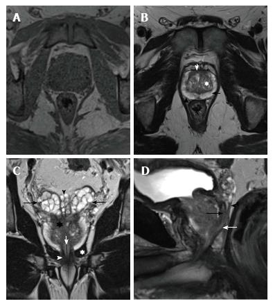
Figure 1 Normal prostate anatomy on magnetic resonance imaging - T1 and T2-weighted images.
A: T1-weighted axial image of a normal prostate: Homogeneous gland, isointense to the adjacent pelvic muscles. It is not possible to differentiate any anatomical detail; B: Axial image of the prostate shows peripheral zone (PZ - black arrow) as a hyperintense area with a U-shape. The transitional zone (TZ - white asterisk) has a hypointense multinodular pattern (“organized chaos”). Anterior fibromuscular stroma (white arrow) is seen as a hypointense area anterior to the TZ and medial to both anterior PZ horns. The “capsule” (arrowhead) is seen as a hypointense rim surrounding the gland; C: Coronal image shows the hyperintense seminal vesicles located cephalad to the base of the prostate (black arrows). Ampulla of vas deferens (VD - black arrowhead) can be seen as paired structures medial to both seminal vesicles (SV). Prostate central zone (asterisk) is seen as a hypointense area located in the base of the prostate gland. Urethra (white arrow), levator ani muscle (white asterisk) and external sphincter (white arrowhead) are also shown; D: Sagittal image shows the vas deferens (black arrow), seminal vesicles (arrowhead) and the ejaculatory duct (white arrow).
- Citation: Couñago F, Sancho G, Catalá V, Hernández D, Recio M, Montemuiño S, Hernández JA, Maldonado A, del Cerro E. Magnetic resonance imaging for prostate cancer before radical and salvage radiotherapy: What radiation oncologists need to know. World J Clin Oncol 2017; 8(4): 305-319
- URL: https://www.wjgnet.com/2218-4333/full/v8/i4/305.htm
- DOI: https://dx.doi.org/10.5306/wjco.v8.i4.305


