Copyright
©The Author(s) 2017.
World J Clin Oncol. Jun 10, 2017; 8(3): 289-292
Published online Jun 10, 2017. doi: 10.5306/wjco.v8.i3.289
Published online Jun 10, 2017. doi: 10.5306/wjco.v8.i3.289
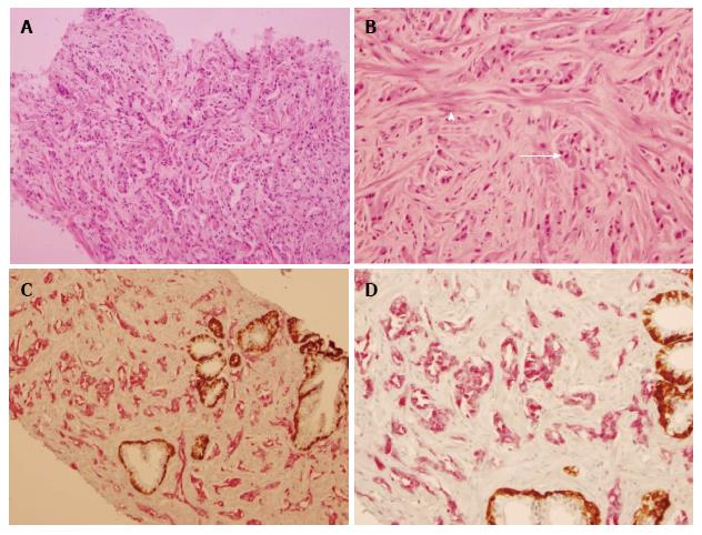
Figure 1 Oncocytic variant of prostatic adenocarcinoma: Hematoxylin and eosin and Immunohistochemical evaluation.
A: Low power view of tumor, showing the tumor cells arranged in glandular and loose epithelial clusters (100 ×); B: High power view of tumor in glandular formations (arrow) and spindled residual prostate myocytes (arrowhead) (200 ×); C: PIN4 immunohistochemical staining identifies tumor (in red) and benign prostatic glands with residual basal cells (in brown) (100 ×); D: PIN4 immunohistochemical staining high power (200 ×) view with tumor (in red) and benign prostatic glands with residual basal cells (in brown).
- Citation: Klairmont MM, Zafar N. Prostatic adenocarcinoma oncocytic variant: Case report and literature review. World J Clin Oncol 2017; 8(3): 289-292
- URL: https://www.wjgnet.com/2218-4333/full/v8/i3/289.htm
- DOI: https://dx.doi.org/10.5306/wjco.v8.i3.289


