Copyright
©The Author(s) 2016.
World J Clin Oncol. Jun 10, 2016; 7(3): 308-320
Published online Jun 10, 2016. doi: 10.5306/wjco.v7.i3.308
Published online Jun 10, 2016. doi: 10.5306/wjco.v7.i3.308
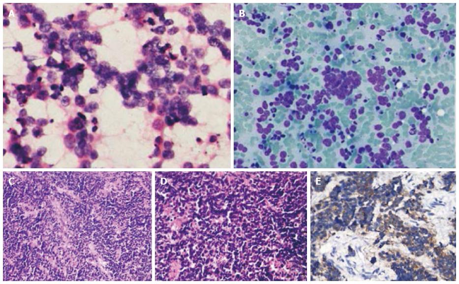
Figure 3 Histopathology and cytology of lymph node samples of illustrative case 2.
A: Microphotograph showing dispersed population of tumor cells along with few loose clusters. The tumor cells with high nuclear cytoplasmic ratio and showing nuclear moulding (MGG × 20 ×); B: Microphotograph showing small tumor cells with high N:C ratio, salt and pepper type chromatin and inconspicuous nucleoli (HE × 40 ×); C: Photomicrographs showing tumour with extensive crushing artefact; D: Tumour cells having hyperchromatic nuclei, scanty cytoplasm and apoptosis; E: Synaptophysin immunostain showing cytoplasmic positivity.
- Citation: Sehgal IS, Kaur H, Dhooria S, Bal A, Gupta N, Behera D, Singh N. Extrapulmonary small cell carcinoma of lymph node: Pooled analysis of all reported cases. World J Clin Oncol 2016; 7(3): 308-320
- URL: https://www.wjgnet.com/2218-4333/full/v7/i3/308.htm
- DOI: https://dx.doi.org/10.5306/wjco.v7.i3.308


