Copyright
©2014 Baishideng Publishing Group Inc.
World J Clin Oncol. Dec 10, 2014; 5(5): 1107-1112
Published online Dec 10, 2014. doi: 10.5306/wjco.v5.i5.1107
Published online Dec 10, 2014. doi: 10.5306/wjco.v5.i5.1107
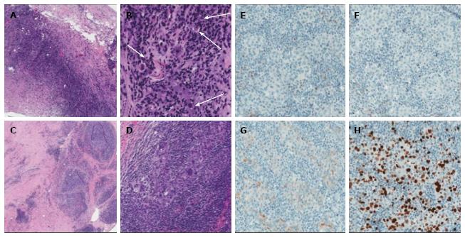
Figure 2 Pathology findings.
A, B: Breast, core needle biopsy: large area of necrosis with chronic inflammation and fibrosis at periphery of lesion, 4X magnification (A); 16X magnification of A showing rare, atypical cells (arrows, B); C, D: Breast, surgical excision: poorly circumscribed lesion adjacent to previous biopsy site, 10X magnification (C); high grade carcinoma with marked pleomorphism and syncytical growth somewhat obscured by marked intra- and peri-turmoral inflammatory infiltrate (D) characteristic of LELC, 10X magnification; E-H: Immunophenotype: ER negative (E), PR negative (F), Her-2/neu 1+ (G), and Ki-67 27% (H). 10X magnification. ER: Estrogen-receptor; PR: Progesterone-receptor.
- Citation: Suzuki I, Chakkabat P, Goicochea L, Campassi C, Chumsri S. Lymphoepithelioma-like carcinoma of the breast presenting as breast abscess. World J Clin Oncol 2014; 5(5): 1107-1112
- URL: https://www.wjgnet.com/2218-4333/full/v5/i5/1107.htm
- DOI: https://dx.doi.org/10.5306/wjco.v5.i5.1107


